Figure 2. ARF1 inhibitor local administration on alkali-induced corneal neovascularization. One week after the alkali burn, the mice
were divided randomly into two groups. The treated group received ARF1 inhibitor for 2 weeks, and the control group received
0.2% HA. A: Representative macroscopic observation of corneal neovascularization (CNV) 3 weeks after alkali injury are shown. B: Representative immunofluorescences of corneal microvessel densities in different treatment group 3 weeks after alkali injury
are shown. C: The distribution of the percentage of CNV in each group are shown here (horizontal line represents the average; *p<0.05
versus the control group).
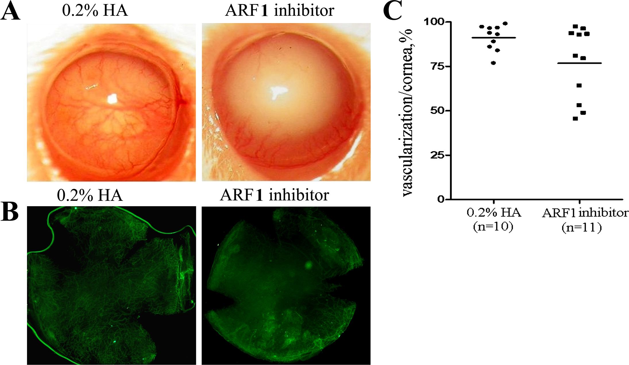
 Figure 2 of
Dai, Mol Vis 2012; 18:2947-2953.
Figure 2 of
Dai, Mol Vis 2012; 18:2947-2953.