Figure 5. Subcellular localization of NHS-A and EPHA2 proteins in human SRA 01/04 and mouse αTN4 lens epithelial cells. Endogenous NHS-A
and EPHA2 proteins were immunolabeled respectively with the anti-NHS and anti-EphA2 antibody. NHS-A localized to the cellular
periphery as punctate strands perpendicular to the cell boundary in both the cell types. EPHA2 showed uniformly speckled localization
in the cytoplasm in SRA 01/04 cells; in αTN4 cells it localized in the cytoplasm as discrete specks; in positive control Madin-Darby
canine kidney (MDCK) cells, it localized to the cellular periphery. The absence of a signal in each cell type upon hybridization
with the secondary antibody alone as a negative control (not shown) proved specificity of localization of each labeled protein.
NHS-A labeling was detected by epifluorescence microscopy using a 40× objective. EPHA2 labeling was detected by confocal microscopy
using a 60× objective and further digital magnification. Representative images from two independent experiments are shown.
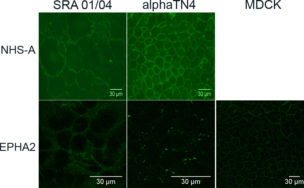
 Figure 5 of
Dave, Mol Vis 2012; 18:2937-2946.
Figure 5 of
Dave, Mol Vis 2012; 18:2937-2946.