Figure 1. Expression of cellular junctions associated and cytoskeletal proteins in human SRA 01/04 and mouse αTN4 lens epithelial cells.
Western blotting was performed on protein lysates of SRA 01/04 and αTN4 cells using the (A) anti-zona occludin-1 (ZO-1), (B) anti-occludin, (C) anti-E-cadherin, and (D) anti-β-actin antibodies. Protein lysate of MCF-7 human breast cancer cells was used as a positive control in B, C, and D. A: A band of ~220 kilo Daltons (kDa) of the expected size of ZO-1 was detected in each cell line. B: A ~60 kDa band corresponding to the expected size of occludin was detected in SRA 01/04 and the positive control MCF-7 cells.
A very faint band of similar size was observed in αTN4 cells. The <60 kDa bands in SRA 01/04 and MCF-7 cells likely represent
occludin isoforms and >60 kDa bands in SRA 01/04 cells likely represent nondenatured protein. C: A ~100 kDa band corresponding to the expected size of the full-length E-cadherin was detected in the positive control MCF-7
cells, but no signal was detected in SRA 01/04 or αTN4 cells. The <100 kDa bands in MCF-7 cells are likely E-cadherin cleavage
products. D: A ~42 kDa band of the expected size of β-actin was detected in all the cell lines. Molecular masses of protein standards
are indicated in kDa.
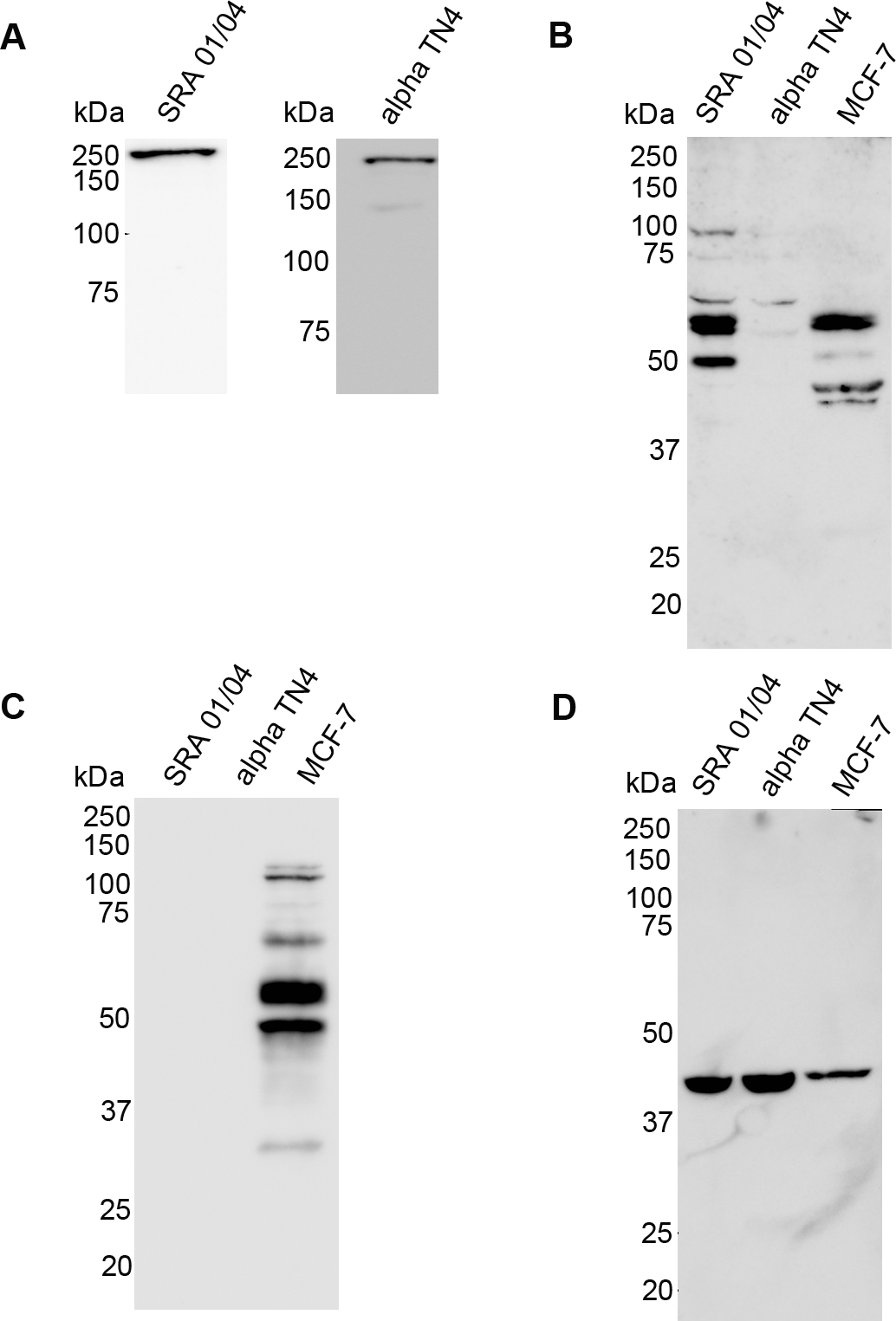
 Figure 1 of
Dave, Mol Vis 2012; 18:2937-2946.
Figure 1 of
Dave, Mol Vis 2012; 18:2937-2946.