Figure 7. Primary mouse retinal ganglion cells isolated by immunopanning-magnetic separation. Glial fibrillary acidic protein–labeled
cells were detected with green fluorescence (B). Syntaxin 1-labeled cells were detected with red fluorescence (C). 4',6-diamidino-2-phenylindole nuclear staining was detected with blue fluorescence (D). Merge image was constructed (A). Scale bars: 100 μm.
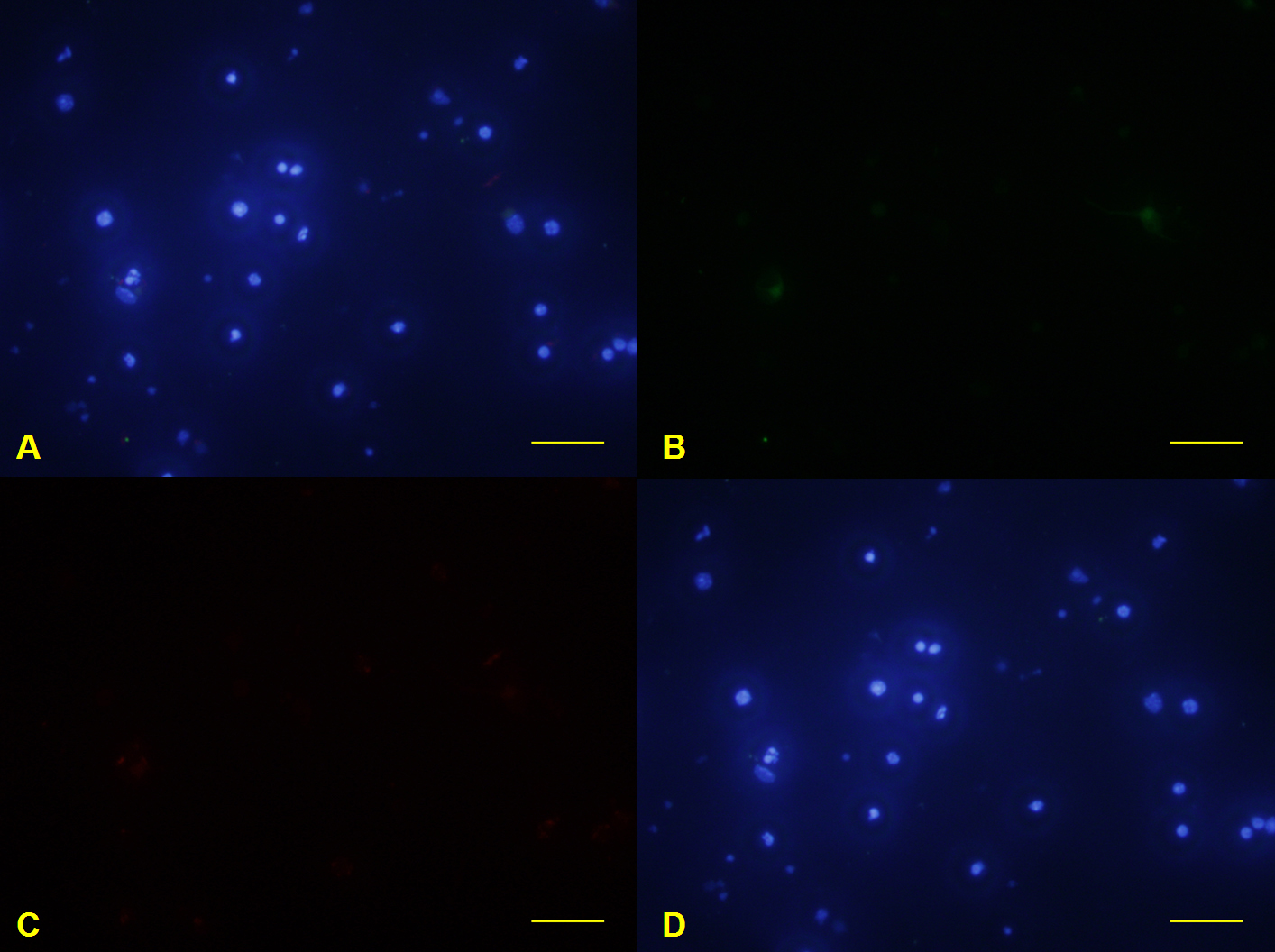
 Figure 7 of
Hong, Mol Vis 2012; 18:2922-2930.
Figure 7 of
Hong, Mol Vis 2012; 18:2922-2930.