Figure 2. Optical coherence tomography and fundus photographs of affected individuals. A: Optical coherence tomography (OCT) of the right eye of individual IV:3 at the age of 38 years shows macular thinning and
loss of foveal contour. B: OCT of the right eye of individual IV:2 at the age of 36 years shows severe thinning of the macula, with irregular retinal
pigment epithelium (RPE). C: Fundus photograph of individual IV:3 at the age of 38 years demonstrating pink optic disc and moderately attenuated retinal
vessels. Macular involvement is indicated by the lack of macular reflex, and by macular atrophy. Pigmentary changes (beaten
bronze like) are observed in the macular region. D: Fundus photograph of retinal periphery in individual IV:3 demonstrating bone spicule-like pigmentation deposits and punctate
salt- and pepper-like appearance.
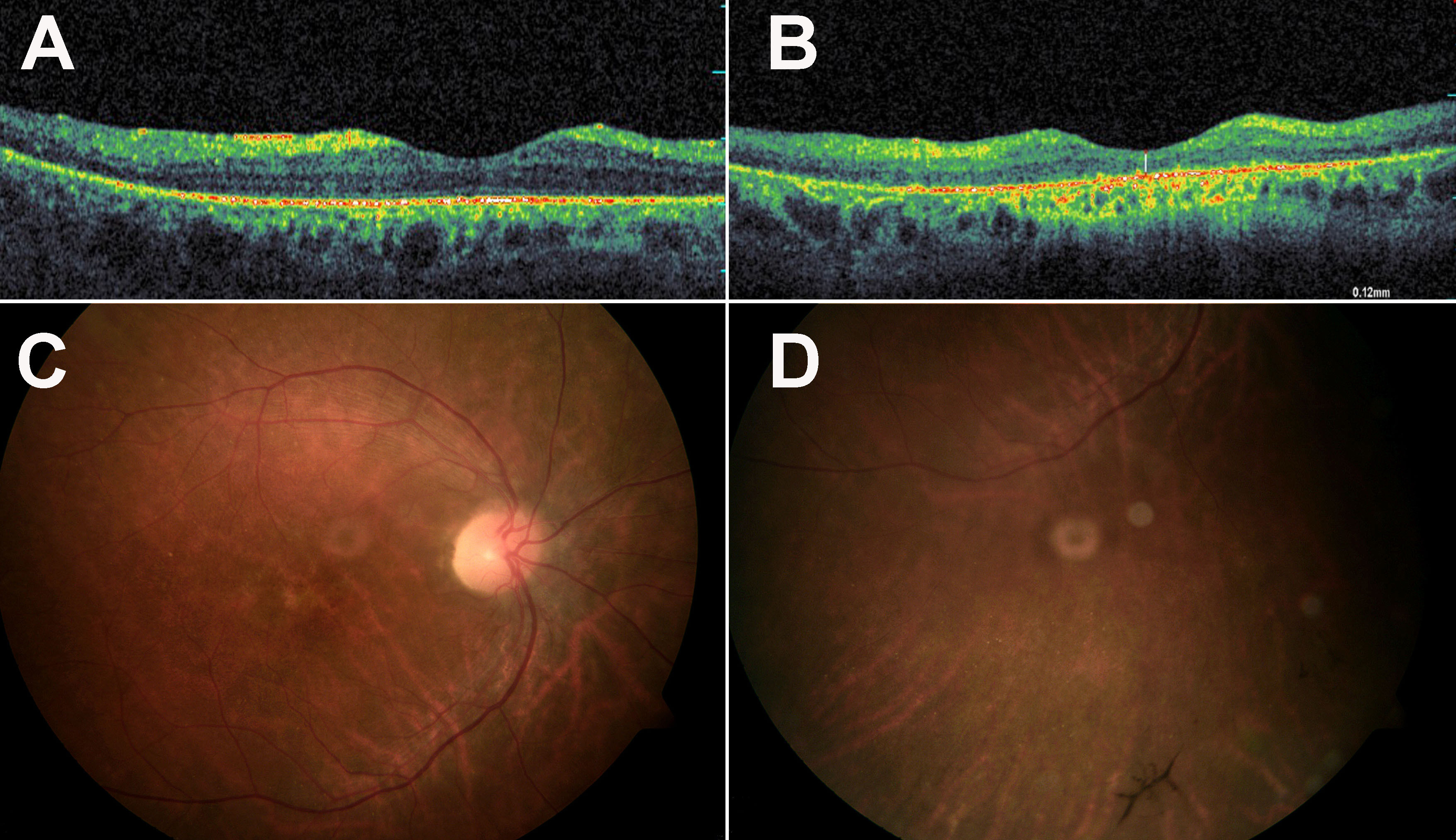
 Figure 2 of
Cohen, Mol Vis 2012; 18:2915-2921.
Figure 2 of
Cohen, Mol Vis 2012; 18:2915-2921.