Figure 5. Corneas cultured in fully immersed conditions. For each condition (E=control with the epithelium and limbus intact; WE=wounded
without prior removal of the epithelium and limbus; WET=as WE but with TGF-β1 added at 10 ng/ml; WT=wounded after prior removal
of the epithelium and limbus plus TGF-β1 added at 10 ng/ml), keratocyte activation (see arrows) and stromal disorganization
were assessed with HPS staining (scale bars: 100 µm (row 1), 50 µm (row 2)), expression of α-smooth muscle actin (SMA), fibronectin
(FN), tenascin C (TNC), and thrombospondin-1 (TSP) with immunofluorescence (scale bars=100 µm), and corneal opacity (OP) by
the ability to read Arial 8 characters (number of opaque corneas / total number of corneas examined for each condition). Each
figure shows the wound region only, except the control E, which shows the central part of the cornea.
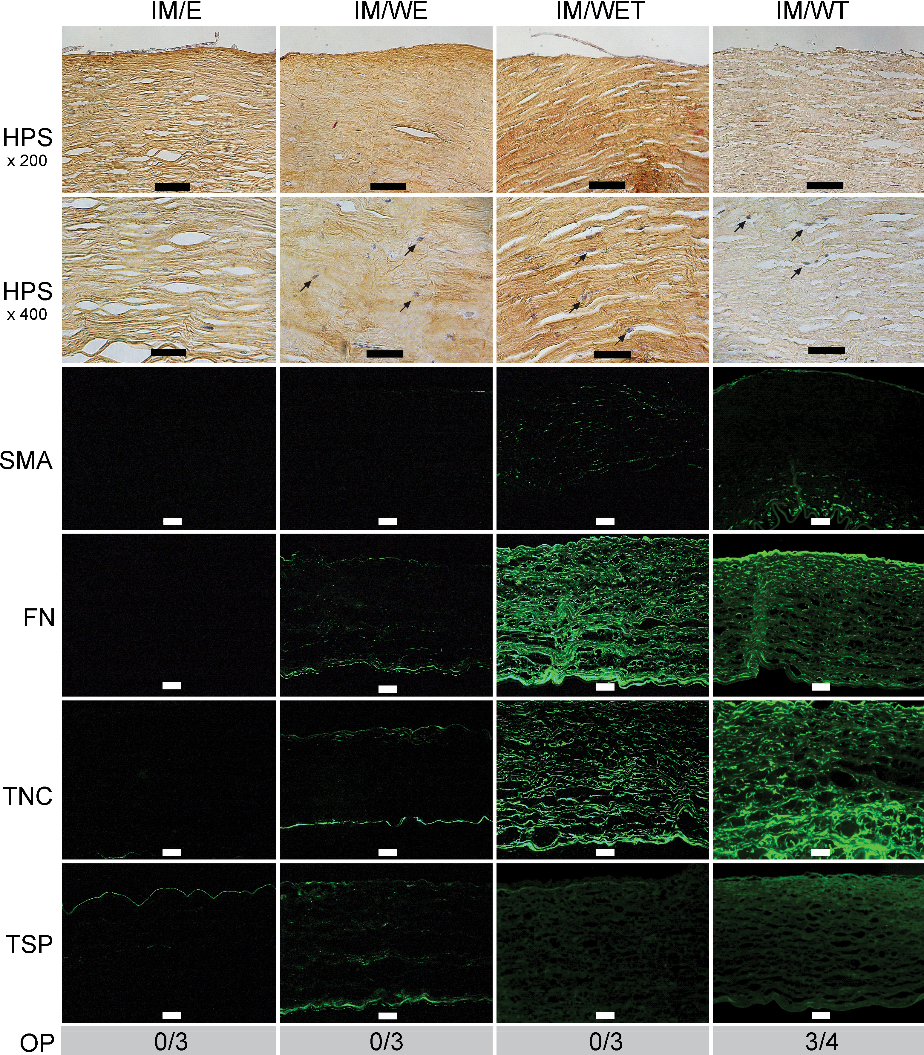
 Figure 5 of
Janin-Manificat, Mol Vis 2012; 18:2896-2908.
Figure 5 of
Janin-Manificat, Mol Vis 2012; 18:2896-2908.