Figure 4. Assessment of corneal opacity. While corneas wounded without prior removal of the epithelium and the limbus (AL/WE, IM/WE)
are transparent, corneas wounded after prior removal of the epithelium and limbus and cultured with added TGF-β1 (AL/WT, IM/WT)
are opaque. Note that the latter corneas are smaller in diameter due to the total elimination of the limbus by trepanation
(8 mm diameter trepan). Scale bars=5 mm.
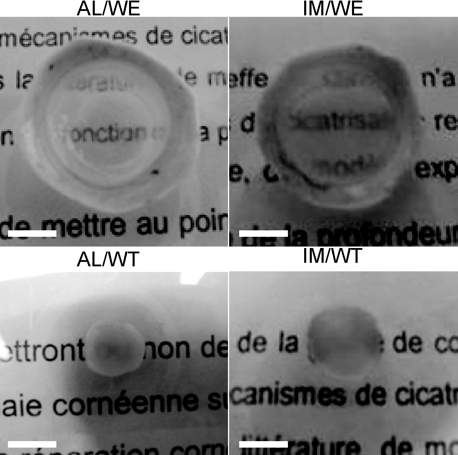
 Figure 4 of
Janin-Manificat, Mol Vis 2012; 18:2896-2908.
Figure 4 of
Janin-Manificat, Mol Vis 2012; 18:2896-2908.