Figure 1. Conditions tested in this study. A: Details of procedures performed before subsequent culture, including removal of the epithelium and the outer part of the
cornea including the limbus, leaving the epithelial basement membrane intact, and wounding using an excimer laser. B: Diagrammatic representation of the different culture conditions used, at the air-liquid interface (AL) and fully immersed
in culture medium (IM). Conditions include non-wounded controls with the epithelium and limbus intact (represented by E) and
wounded corneas (W) with and without prior removal of the epithelium and limbus, and with and without TGF-β1 (T) added at
a concentration of 10 ng/ml.
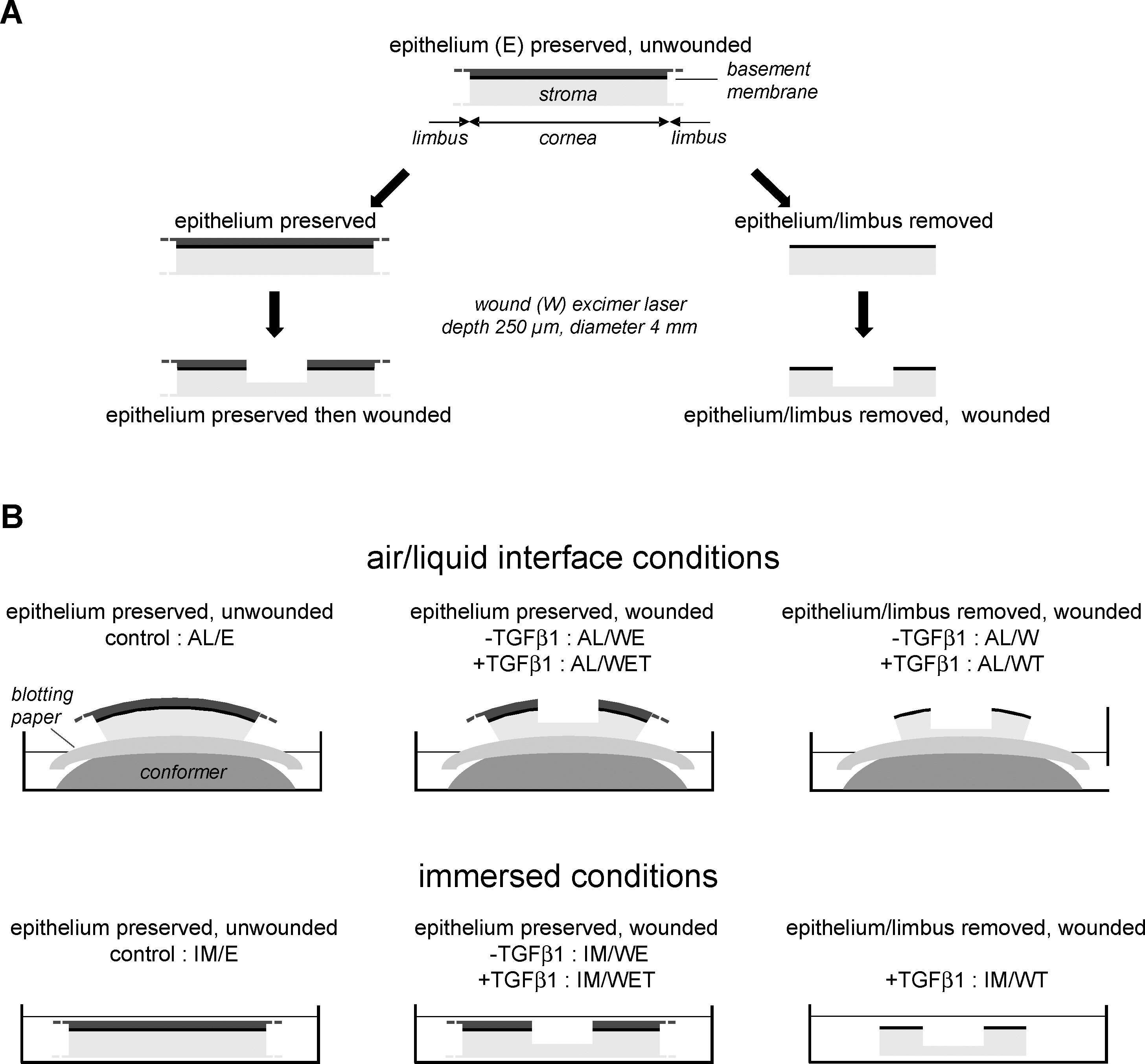
 Figure 1 of
Janin-Manificat, Mol Vis 2012; 18:2896-2908.
Figure 1 of
Janin-Manificat, Mol Vis 2012; 18:2896-2908.