Figure 4. Expression of human embryonic stem cell markers in human Tenon’s capsule fibroblast derived induced pluripotent stem cells.
A: Confocal immunofluorescent images of representative human Tenon’s capsule fibroblast derived induced pluripotent stem cells
(TiPS) clones stained with the human embryonic stem (hES) markers SSEA-4 (red), TRA-1–60 (green), Nanog (red), and Oct-4 (red).
Nuclei were stained with DAPI (blue). hESCs were used as a control. Scale bars=50 μm. B: Flow cytometry analysis of the equivalent SSEA-4 expression levels in TiPS and hESCs. 99.5% and 99.7% SSEA-4 + cells (in
the Q4 area), respectively, whereas 0.3% in HTFs (p<0.001).
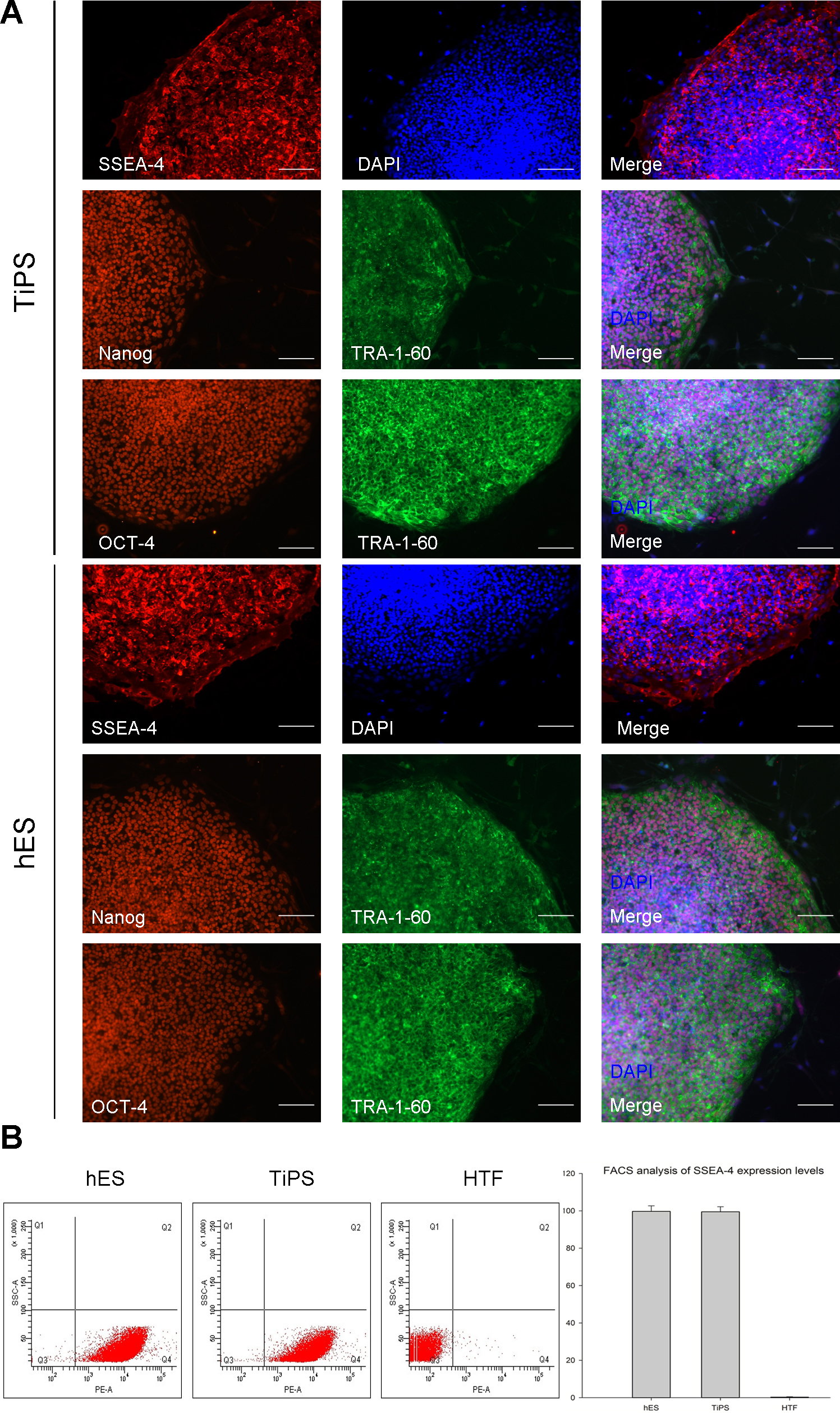
 Figure 4 of
Deng, Mol Vis 2012; 18:2871-2881.
Figure 4 of
Deng, Mol Vis 2012; 18:2871-2881.