Figure 3. Generation of induced pluripotent stem cells from human Tenon’s capsule fibroblasts. A: Immunofluorescent image of human Tenon’s capsule fibroblasts (HTFs) 24 h after infection with a control green fluorescent
protein (GFP) vector. An infection efficiency of nearly 100% was observed in cells infected with the control GFP vector. Scale
bar=200 μm B: Phase contrast image of mesenchymal-epithelial transition (MET). During the early stages of reprogramming, fibroblasts underwent
a morphological transformation from a mesenchymal to an epithelial-like morphology. Scale bar=50 μm and 100 μm, respectively.
C: Phase contrast image of granulated and human embryonic stem cell (hESC)-like colonies. The typical hESC-like colony was
flat, with well defined phase-bright borders. Scale bar=200 μm. D: Phase contrast images of an established induced pluripotent stem (iPS) cell line at passage P 13 and a control hES cell
line at passage P 77. Both include typical honeycomb cells with a high nucleus-to-cytoplasm ratio and prominent nucleoli.
Scale bar=200 μm and 100 μm, respectively.
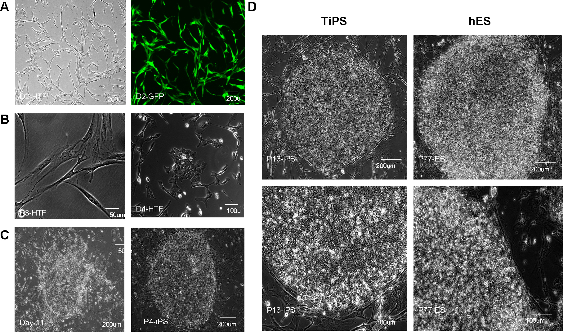
 Figure 3 of
Deng, Mol Vis 2012; 18:2871-2881.
Figure 3 of
Deng, Mol Vis 2012; 18:2871-2881.