Figure 2. Extraction and characterization of human Tenon’s capsule fibroblasts. A: Phase contrast images of human Tenon’s capsule fibroblasts (HTFs). Generally flat, elongated, spindle-shaped cells climb
out of the tissue approximately 1 week after seeding. Scale bar=200 μm B: Confocal immunofluorescent images of HTFs. Expression of fibronectin and vimentin. Scale bar=20 μm.
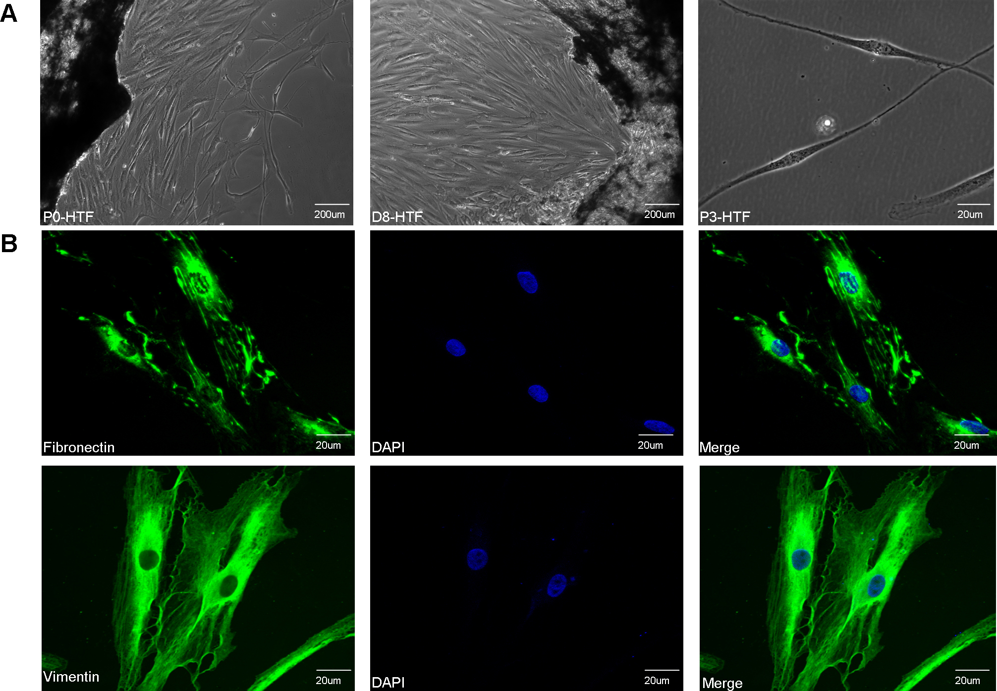
 Figure 2 of
Deng, Mol Vis 2012; 18:2871-2881.
Figure 2 of
Deng, Mol Vis 2012; 18:2871-2881.