Figure 6. The effect of 15-LOX-1 overexpression on preretinal neovascularization in mice with oxygen-induced ischemic retinopathy. The
experiments were divided into the oxygen-induced retinopathy treated with adenoviral-15-lipoxygenase-1 group (the 15-LOX-1
group), the untreated oxygen-induced retinopathy group (the OIR group), the oxygen-induced retinopathy treated with adenoviral-green
fluorescence protein group (the GFP group) and the normal control group (the normal group), with eight mice per experimental
group. At postnatal day 17, all mice were fixed, sectioned, and stained with hematoxylin and eosin. Preretinal neovascular
cells were counted on eight non-continuous sections per eye and averaged. The images were representatives the retinal sections
from the eyes of 15-LOX-1 group (A), OIR group (B), GFP group (C) and Normal group (D). Arrows indicate preretinal neovascular cells. The magnification of A-D is 200×. Scale bars=50 um. E: The average numbers of preretinal neovascular cells (means±SD, n=8) of the 15-LOX-1 group mice were compared with the OIR
group, GFP group, and normal group mice using one-way ANOVA. 15-LOX-1 group versus OIR group *p<0.05, n=8; 15-LOX-1 group
versus GFP group **p<0.05, n=8.
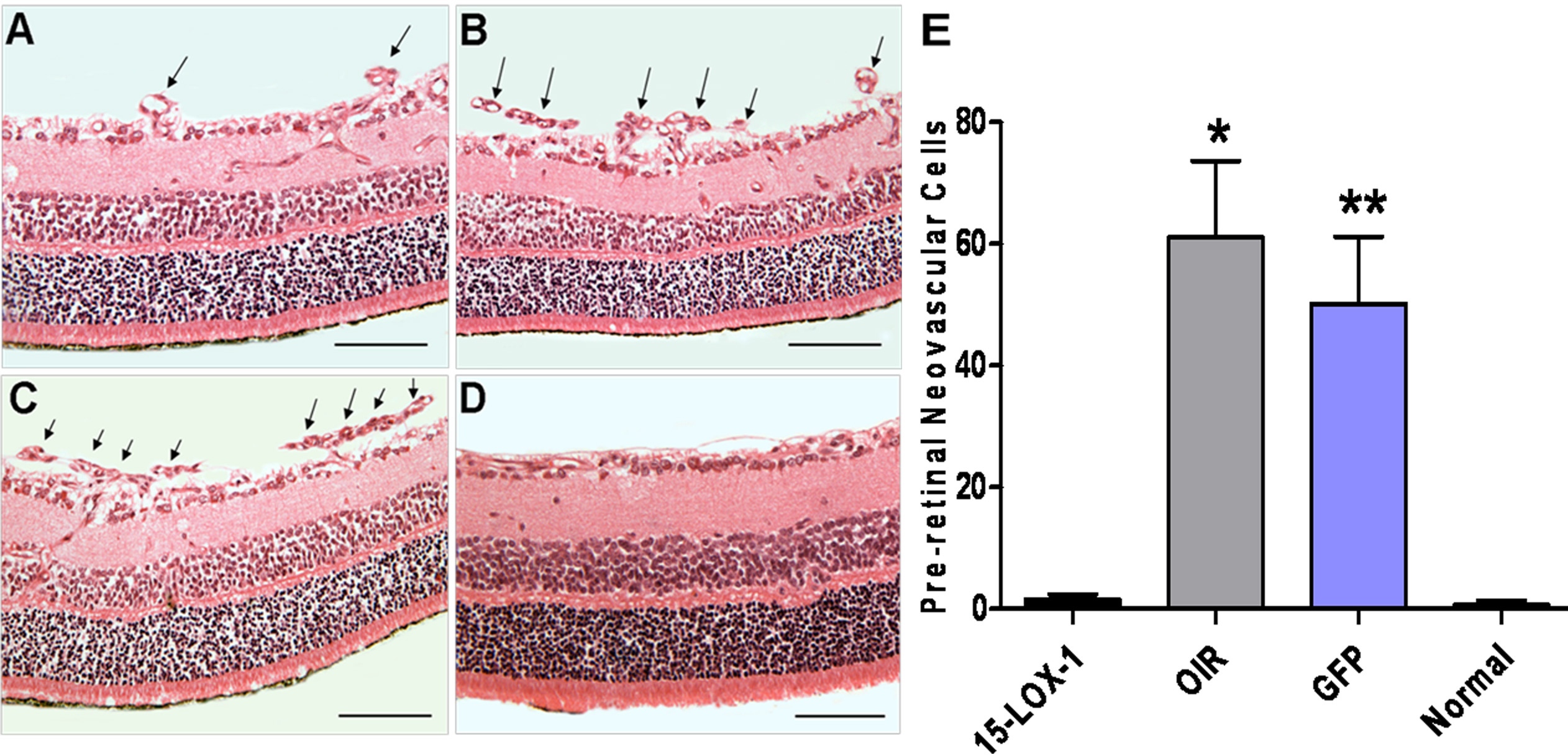
 Figure 6 of
Li, Mol Vis 2012; 18:2847-2859.
Figure 6 of
Li, Mol Vis 2012; 18:2847-2859.