Figure 5. Immunohistochemistry of
cultured BCECs on PCL 25 blend membrane. A: The
expressions of cytoskeleton actin filament of BCECs at day 7 by
confocal microscope. B: Correspondingly, the expressions
of ZO-1 (green) at the margin of cells after 7 days. Scale
bar=50 µm.
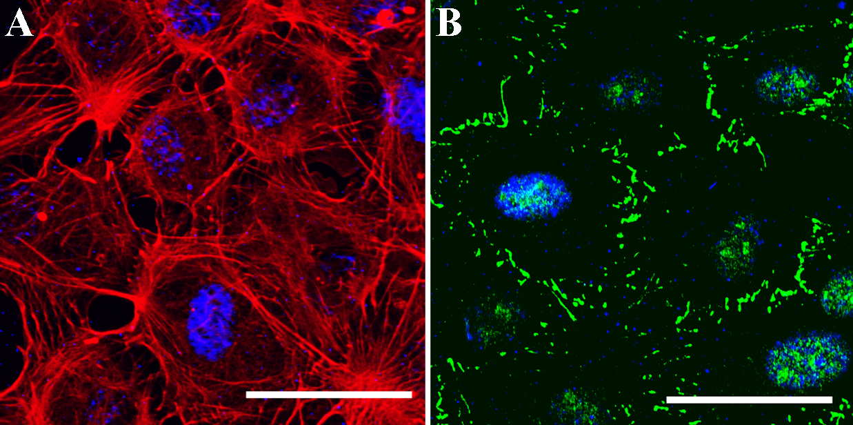
 Figure 5
of Wang, Mol Vis 2012; 18:255-264.
Figure 5
of Wang, Mol Vis 2012; 18:255-264.