Figure 4. Prolonged calcineurin activation caused optic nerve damage. A and B: Sample optic nerve images showed healthy and degenerated axons seven weeks (A) and 16 weeks (B) after AAV injection. Arrows
point at degenerated retinal ganglion cell axons. Scale bar is 10 µm. C and D: The optic nerve degeneration was assessed by optic nerve grading using the Morrison standard. Higher grades were assigned
to more damaged optic nerves. The optic nerve grades of the full-length calcineurin (CaNwt)-expressing eyes and the cleaved
calcineurin (CaNCA)-expressing eyes were normalized to their contralateral control EGFP-expressing eyes. The optic nerves
in the CaNCA-expressing eyes were not different from the CaNwt-expressing eyes seven weeks after AAV injection (C), but were more damaged 16 weeks after AAV injection (D; two-tailed unpaired Student t test).
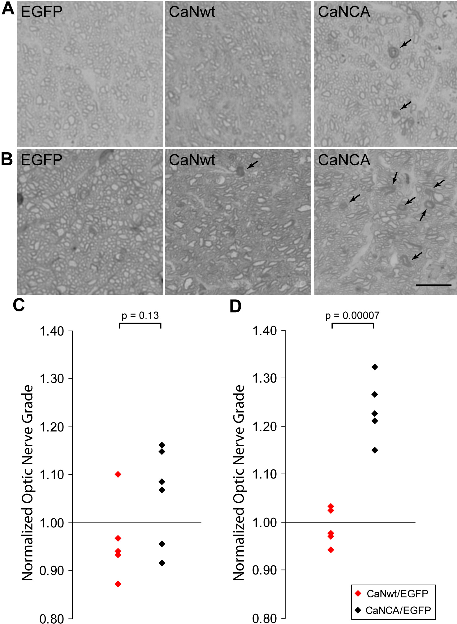
 Figure 4 of
Qu, Mol Vis 2012; 18:2828-2838.
Figure 4 of
Qu, Mol Vis 2012; 18:2828-2838.