Figure 7. NMDAR1 colocalizes with OPN4 in retinal ganglion cells. Shown are representative photomicrographs taken from retinal flat
mounts of untreated wild type mice stained for A, D: NMDAR1 (green) and B, E: OPN4 (red) at A-C: lower and D-F: higher magnification. C, F: Images shown in panels A and B, and in panels D and E, respectively, are merged. Focal plane was at the ganglion cell layer. Scale bars are A-C: 100 µm and D-F: 50 µm.
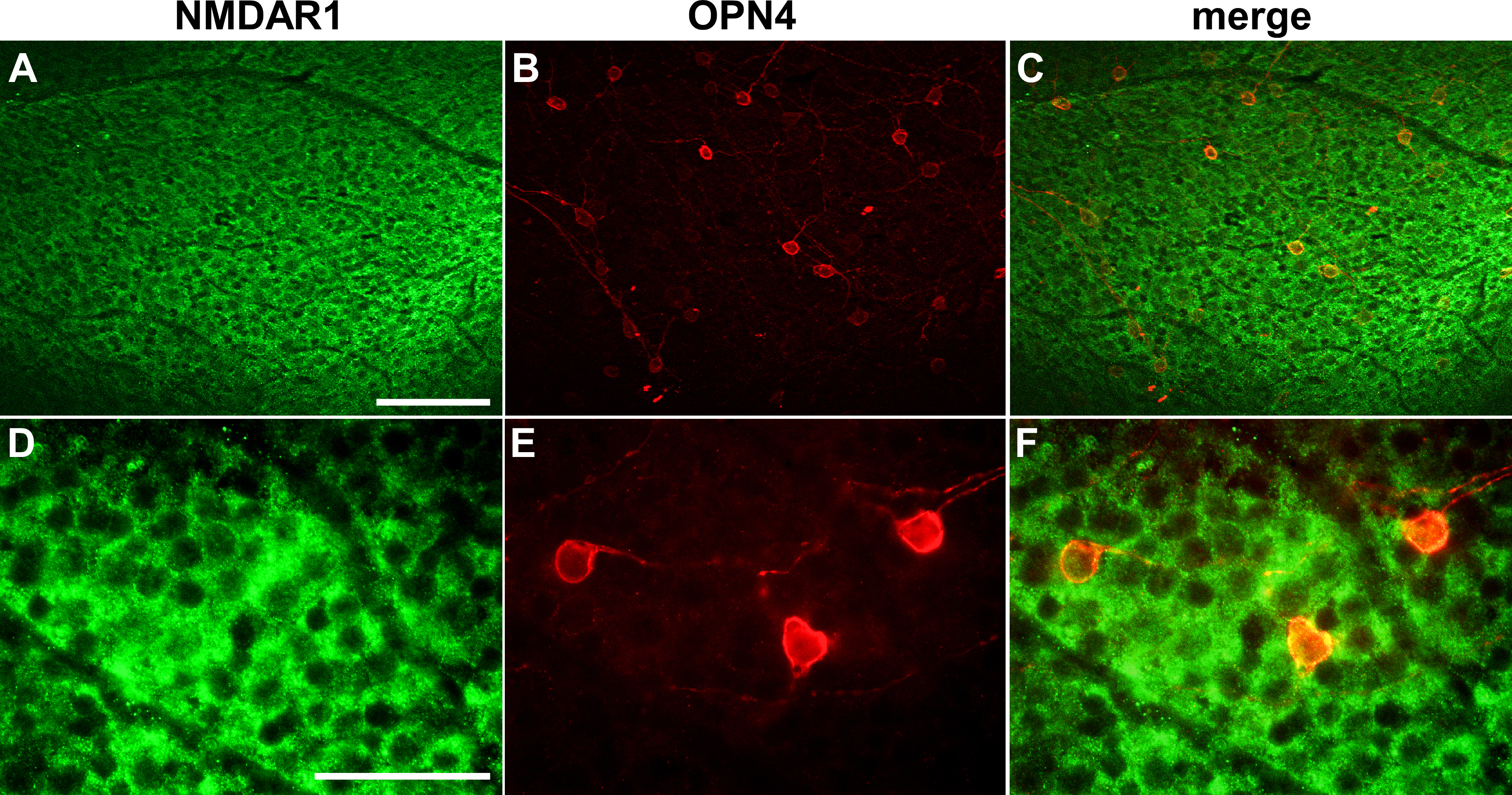
 Figure 7 of
DeParis, Mol Vis 2012; 18:2814-2827.
Figure 7 of
DeParis, Mol Vis 2012; 18:2814-2827.