Figure 1. Cells in the ganglion cell layer are lost after intravitreal injection of N-methyl-D-aspartic acid. A-C: Shown are representative photomicrographs of retinal sections from wild type mice 6 days after intravitreal injection of
A: N-methyl-D-aspartic acid (NMDA) or B: phosphate buffered saline (PBS). C: Untreated retinas served as controls. D-F: Shown are representative photomicrographs of immunfluorescent stainings for BRN3A (yellow) and DAPI (blue) in semithin sagittal
sections through retinas 6 days after injection of D: NMDA or E: PBS. F: Uninjected eyes served as controls. Note the non-specific staining of blood vessels by the secondary antibody, especially
in the outer plexiform layer (OPL), inner nuclear layer (INL), and inner plexiform layer (IPL). G: Cell bodies in the ganglion cell layer (GCL) were quantified at 6 days after intravitreal injection of NMDA (black bar)
or PBS (grey bar), and in untreated eyes (white bar). Shown are means±SD (n=3) for all treatments and analyses. Scale bars
were A-C: 20 µm, D-F: 50 µm. **: p<0.01. A one-way ANOVA with Bonferroni post hoc test was used to test statistical significance.
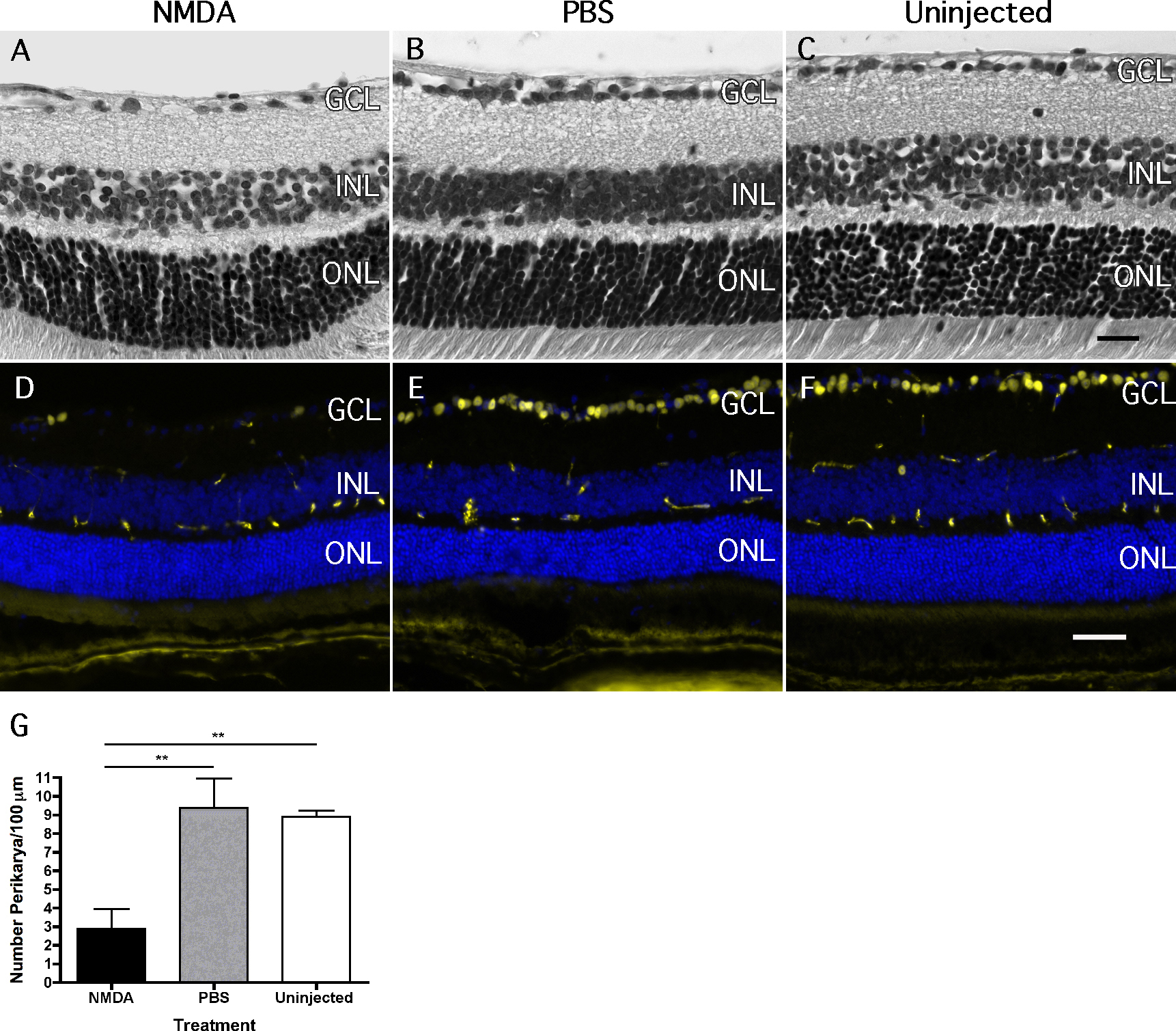
 Figure 1 of
DeParis, Mol Vis 2012; 18:2814-2827.
Figure 1 of
DeParis, Mol Vis 2012; 18:2814-2827.