Figure 1. The RB116 tumor is shown with accompanying histology. A: The RB116 tumor is shown after enucleation. The tumor fills a significant portion of the globe. B: An RB116 tumor section stained with hematoxylin and eosin is magnified 12.5X. The tumor is extensive and displaces the normal
retina. C: A 400X view of the RB116 tumor reveals Flexner-Wintersteiner rosettes (arrows). The scale bar in panel B is one millimeter. The scale bar in panel C is 25 microns.
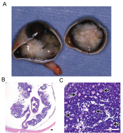
 Figure 1 of
Bejjani, Mol Vis 2012; 18:2805-2813.
Figure 1 of
Bejjani, Mol Vis 2012; 18:2805-2813.