Figure 7. Uptake of aptamer-Dox conjugate on the Y79 cell line post 2 h addition in vitro. Free Dox, EpDT3-Dox, and Scr-EpDT3-Dox were
incubated with Y79 cells for 2 h and observed under an AxioObserver microscope. A, B, C, and D: phase images of Y79 cells unstained, treated with free Dox, Scr-EpDT3-Dox or Scr-EpDT3-Dox and the respective right panel
represents the merged fluorescence image.
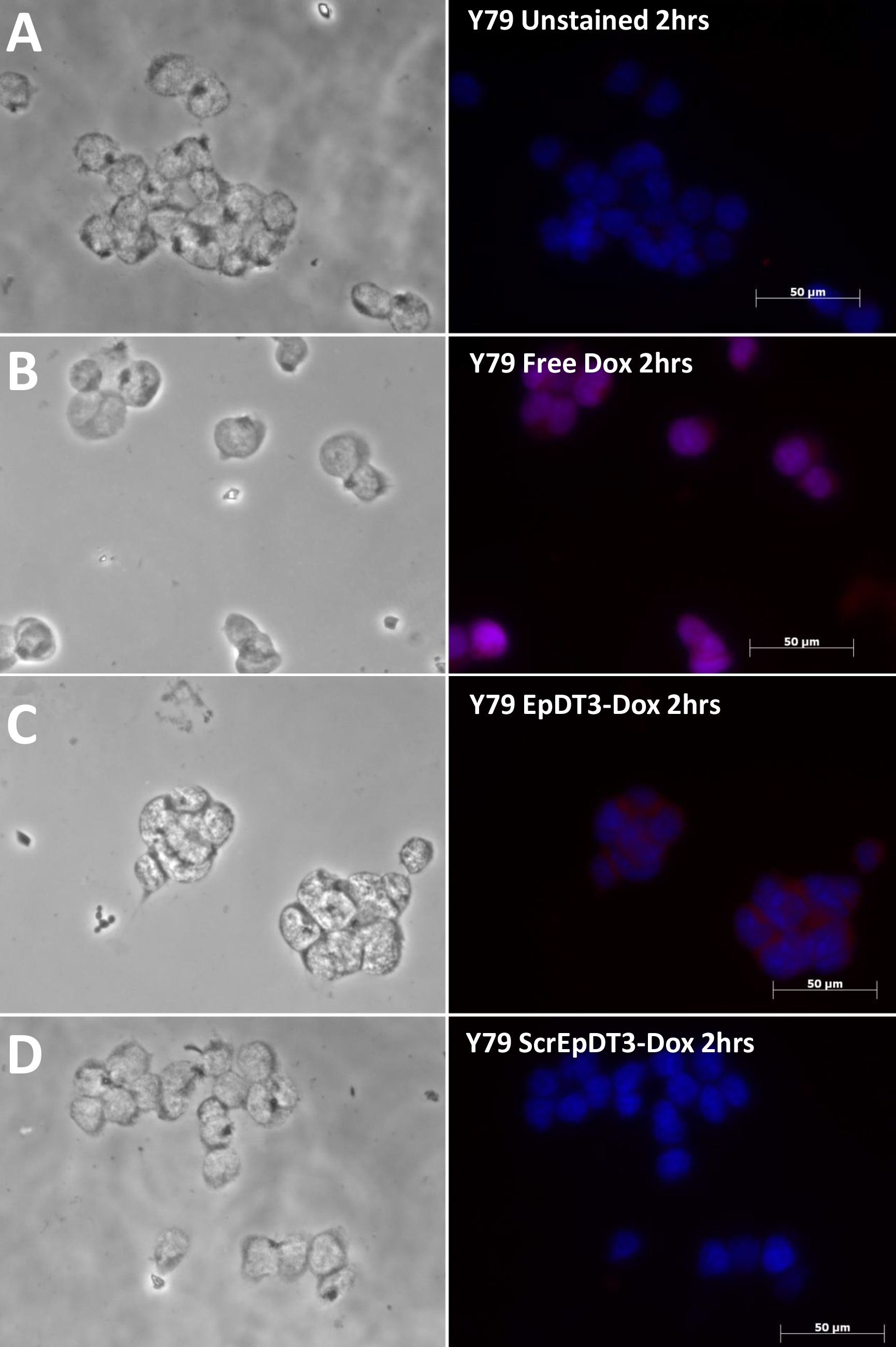
 Figure 7 of
Subramanian, Mol Vis 2012; 18:2783-2795.
Figure 7 of
Subramanian, Mol Vis 2012; 18:2783-2795.