Figure 4. EpCAM aptamer structure and doxorubicin conjugation. Structure prediction was performed using the RNA Structure program. A: Hairpin structure of the EpCAM aptamer with the predicted site for the doxorubicin intercalation represented as a red star.
B: Fluorescence spectra of doxorubicin (3.0 μm) with increasing molar ratios of the aptamer (from top to bottom: 0, 0.01, 0.1,
1, 3, 5, and 7 equivalents) in conjugation buffer.
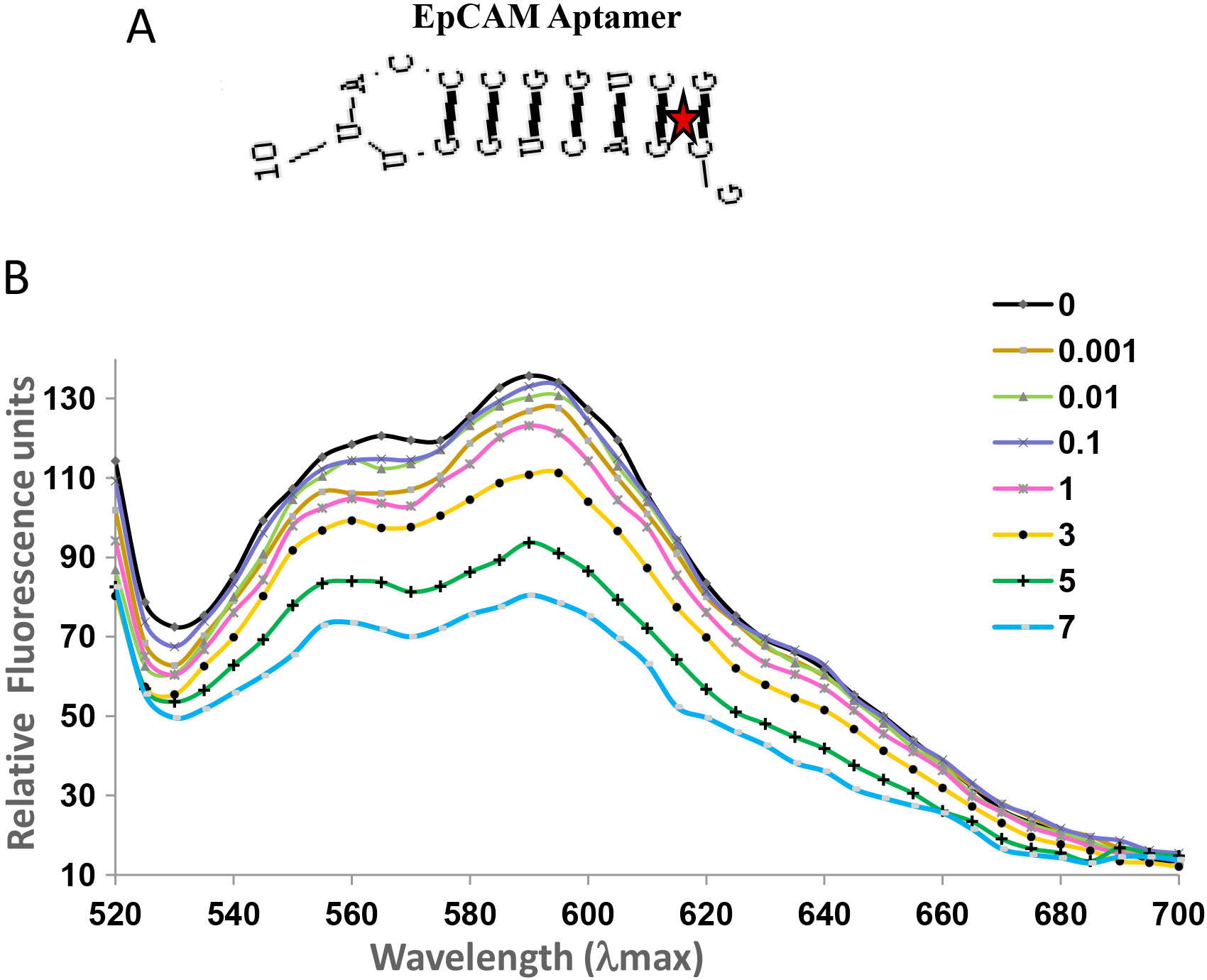
 Figure 4 of
Subramanian, Mol Vis 2012; 18:2783-2795.
Figure 4 of
Subramanian, Mol Vis 2012; 18:2783-2795.