Figure 2. EpCAM aptamer binding on RB primary cells. Fluorescently labeled EpCAM aptamers were incubated with RB cells obtained from
fresh tumor samples and analyzed with flow cytometry. Dead cells were gated out by gating the propidium iodide–positive cells.
A, B, C: Scatter plots show control unstained RB tumor cells, tumor cells stained with EpDT3-FI shifting toward the right, and Scr-EpDT3-FI
showing the least binding. D: Histogram overlay plot showing significant binding of EpDT3-FI to tumor cells compared to Scr-EpDT3-FI.
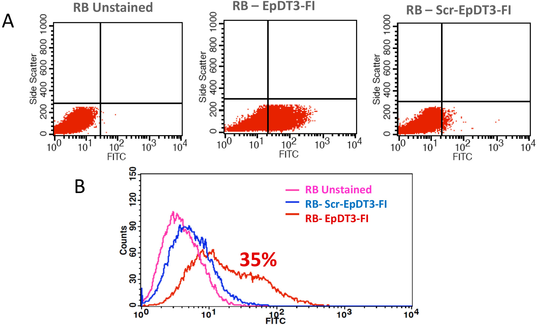
 Figure 2 of
Subramanian, Mol Vis 2012; 18:2783-2795.
Figure 2 of
Subramanian, Mol Vis 2012; 18:2783-2795.