Figure 10. Cell proliferation inhibition by aptamer-Dox conjugates in vitro. WERI-Rb1 cells were seeded 24 h before treatment with free
Dox, and aptamers (EpDT3 and Scr-EpDT3) and aptamer-Dox conjugates (EpDT3-Dox and Scr-EpDT3-Dox; n=3) were added to cells
in serum-free condition for 2 h followed by media change with 10% FBS. Cells were further incubated for 24 h and 48 h, and
an MTT assay was performed. The graph represents the percent viable WERI-Rb1 cells after 24 h and 48 h of treatment. Cytotoxicity
was observed with cells treated with free Dox and EpDT3-Dox.
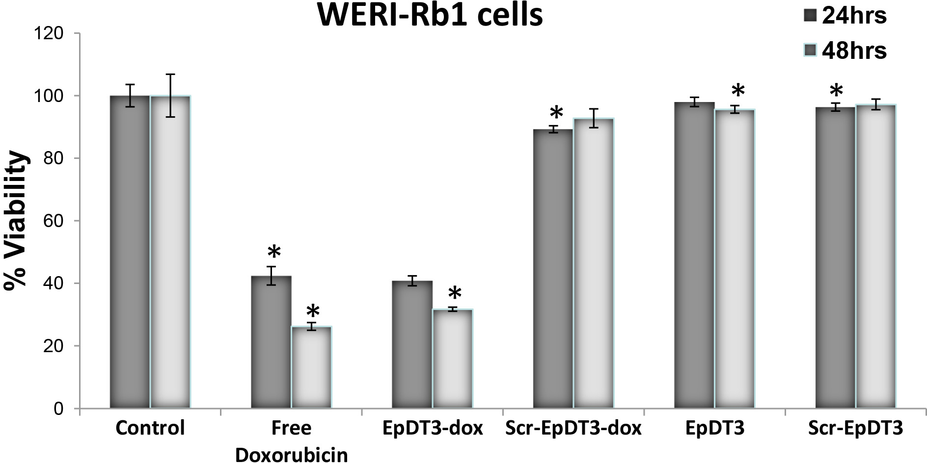
 Figure 10 of
Subramanian, Mol Vis 2012; 18:2783-2795.
Figure 10 of
Subramanian, Mol Vis 2012; 18:2783-2795.