Figure 1. EpCAM expression on cell lines. Y79, WERI-Rb1, and Müller glial cells were stained for EpCAM expression with indirect immunofluorescence
followed by flow cytometry. A, B, C: Y79, WERI-Rb1, and Müller glial cells stained either with secondary antibody alone or primary anti-EpCAM (C-10) antibody,
followed by secondary anti-mouse fluorescein isothiocyanate antibody and flow cytometry. The overlay plot shows the positive
population.
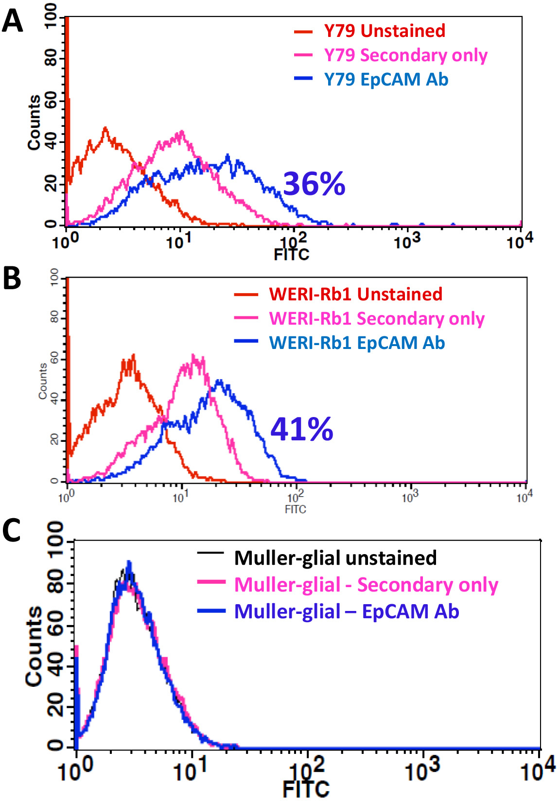
 Figure 1 of
Subramanian, Mol Vis 2012; 18:2783-2795.
Figure 1 of
Subramanian, Mol Vis 2012; 18:2783-2795.