Figure 5. Survivin and proliferating cell nuclear antigen (PCNA) protein expression in the early postnatal mouse lens. A: During early stages of postnatal lens development, western blotting revealed two bands for Survivin at 16.5 kDa and 14 kDa.
The 16.5 kDa band peaked at P7 and diminished in expression thereafter, while the 14 kDa band was expressed at lower levels
at NB and P7 only. Relative expression levels of Survivin, determined with densitometry and normalized to β-actin protein,
revealed statistically significant differences in levels of Survivin protein expression between the calibrator (NB) and the
other stages studied (p<0.05; n=3). B: PCNA protein expression was downregulated between NB and 4 weeks. Relative expression levels of PCNA, determined with densitometry
and normalized to β-actin protein using the scan program, revealed statistically significant differences in levels of PCNA
protein expression between the calibrator NB and the other stages studied (p<0.05; n=3). Representative western blots are
shown. au=arbitrary units.
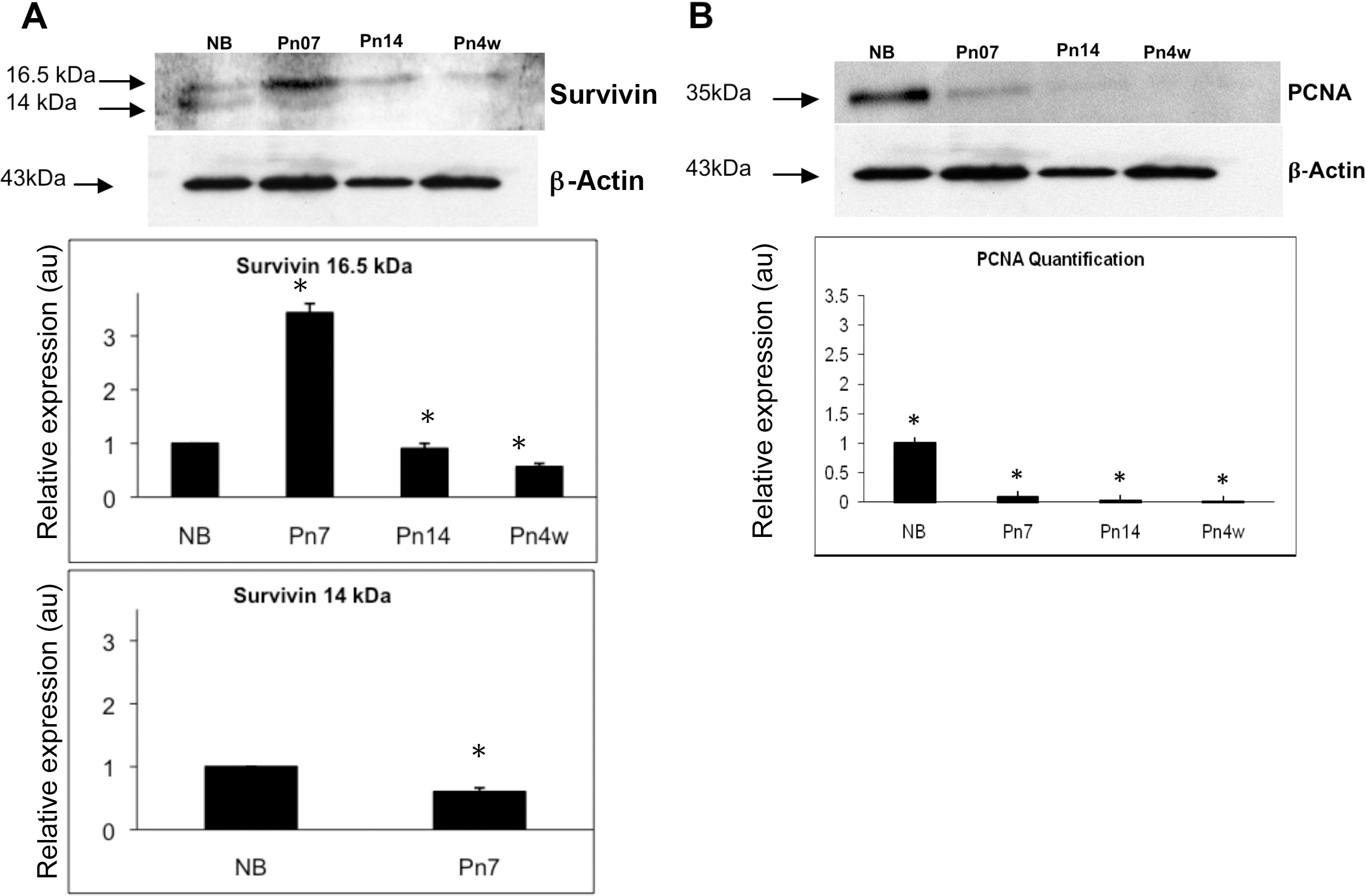
 Figure 5 of
Jarrin, Mol Vis 2012; 18:2758-2769.
Figure 5 of
Jarrin, Mol Vis 2012; 18:2758-2769.