Figure 3. Survivin gene and protein and proliferating cell nuclear antigen (PCNA) protein expression in chick lens epithelial cell primary
cultures. A: Culture of lens dissociated lens epithelial primary cell cultures revealed characteristic morphological changes using phase
contract microscopy as cells progressively differentiated into lens fiber-like cells lentoids (arrows) between day 0 (D0)
and day 8 (D8) of culture. Magnification bars=100 μM. B: Using QPCR, Survivin mRNA expression peaked at D0 and diminished thereafter. Statistical analysis of Survivin expression
using QPCR revealed significant differences in expression levels between all stages studied and the calibrator D0 (*=p<0.05;
n=3). C: Survivin expression peaked at D6 and diminished thereafter; thus, the peak of Survivin protein expression followed the peak
of Survivin RNA expression. The amount of Survivin at each stage of culture was quantified with densitometry using the scan
program and normalized regarding β-actin protein (*=p<0.05; n=3). D: PCNA expression peaked at D6. The amount of PCNA was quantified by densitometry using the scan program and normalized with
respect to the β-actin protein (*=p<0.05; n=3). Chick embryo brain was used as a positive control C+) for Survivin and PCNA
WBs. Representative western blots are shown for Survivin and PCNA. au=arbitrary units.
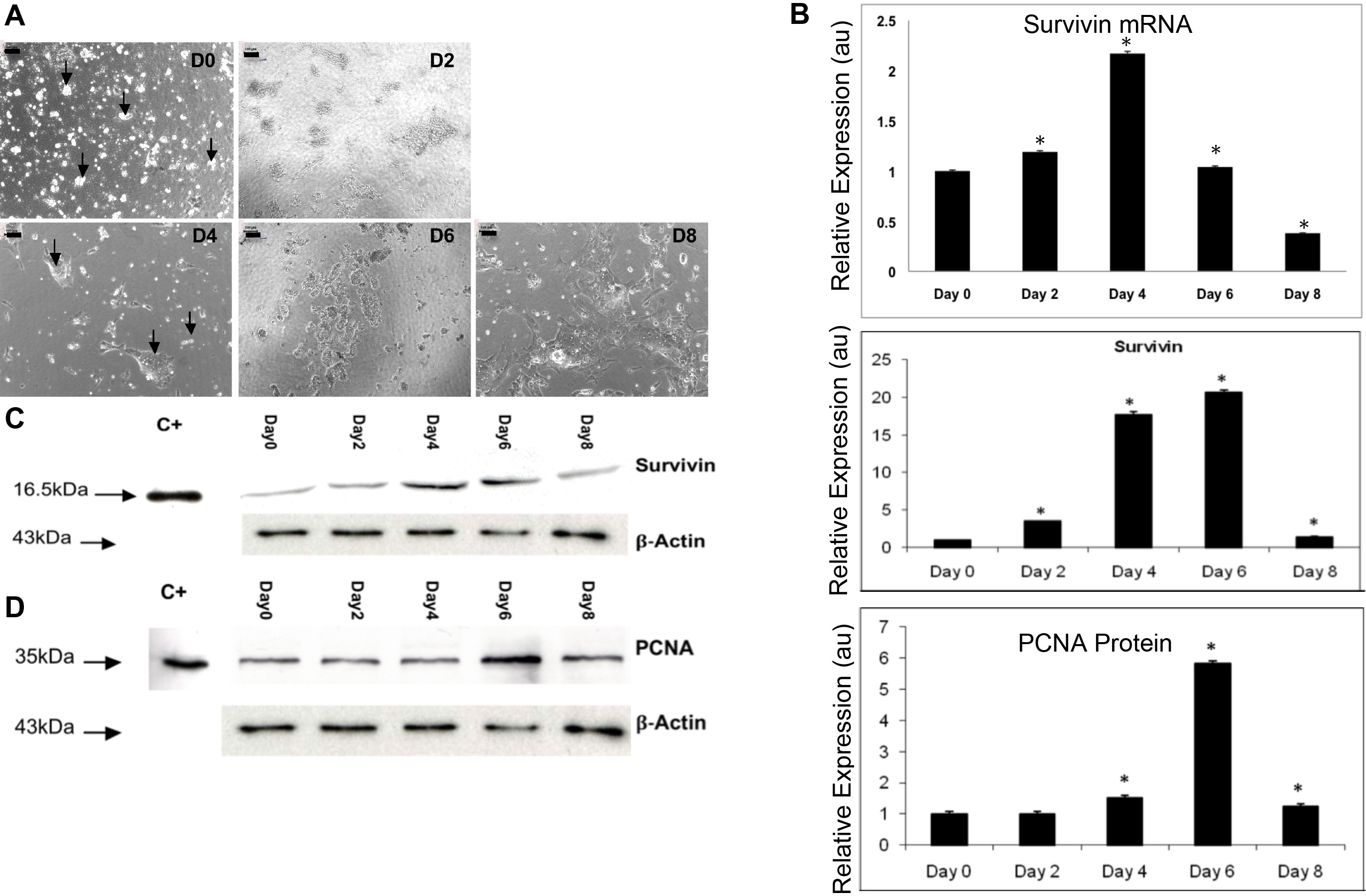
 Figure 3 of
Jarrin, Mol Vis 2012; 18:2758-2769.
Figure 3 of
Jarrin, Mol Vis 2012; 18:2758-2769.