Figure 2. Spatiotemporal immunolocalization of Survivin in the chick embryo lens. A: Immunohistochemistry showed strong expression of Survivin at ED4 in the lens epithelial cells (LECs) and in the lens fiber
cells (LFCs). Survivin staining was associated with the cytoplasm and nuclei in the LECs, but became mainly localized to the
nuclei in the LFCs after passing through the transition zone at the annular pad (AP). B: Higher magnification revealed homogenous Survivin staining in LFC nuclei at ED4. C: In the ED6 lens, Survivin staining was associated with the LECs and the LFC nuclei. D: Higher magnification revealed homogenous Survivin staining in LFC nuclei at ED6. E: At ED12, Survivin staining was associated with marginalized chromatin of pyknotic nuclei of lens fiber cells undergoing
the early stages of denucleation (arrows). E: ED6 negative control lens incubated with rabbit immunoglobulins instead of primary antibody demonstrates the specificity
of Survivin immunostaining. Magnification bars are 100 μM except in E (50 μM).
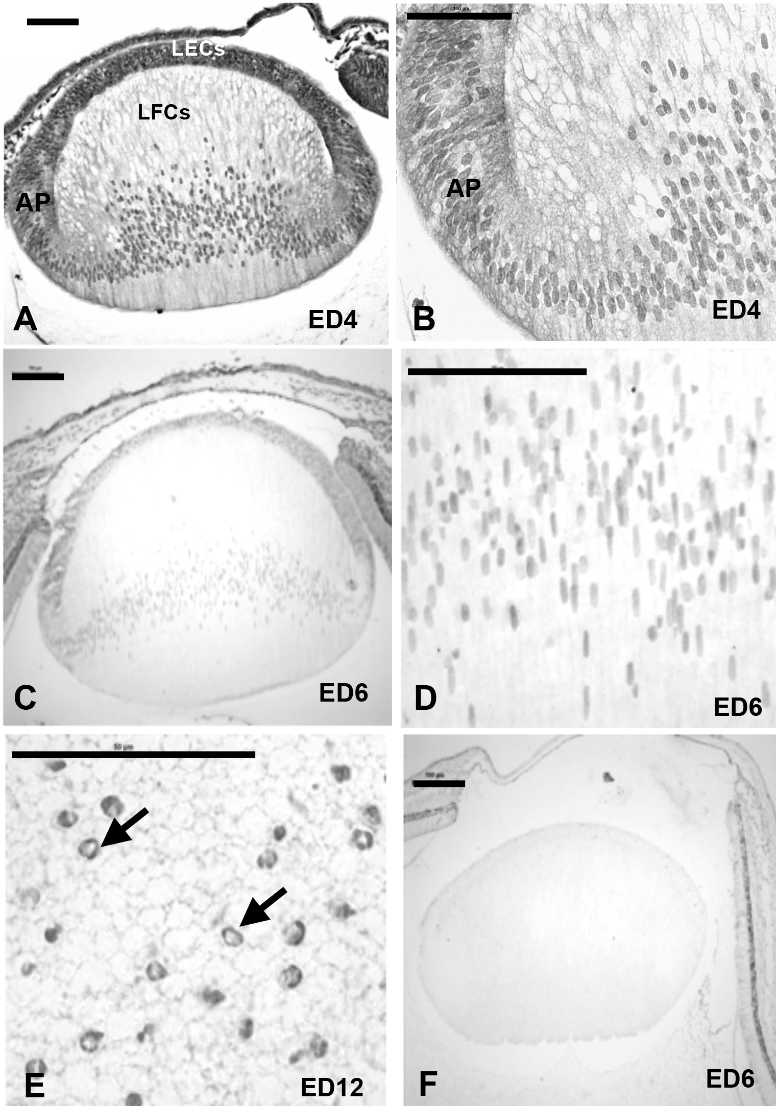
 Figure 2 of
Jarrin, Mol Vis 2012; 18:2758-2769.
Figure 2 of
Jarrin, Mol Vis 2012; 18:2758-2769.