Figure 4. Fundus photographs and OCT of patients J-III-1 (proband) and J-II-2 (proband’s father) showing intrafamilial phenotypic variability.
A, B, C, D: Fibrotic lesion at the posterior pole centered in the macula; the patient received bilateral PDT treatment for CNV (Patient
J-III-1). E, F, G, H: Vitelliruptive stage (Patient J-II-2). OD represents the right eye, OS represents the left eye.
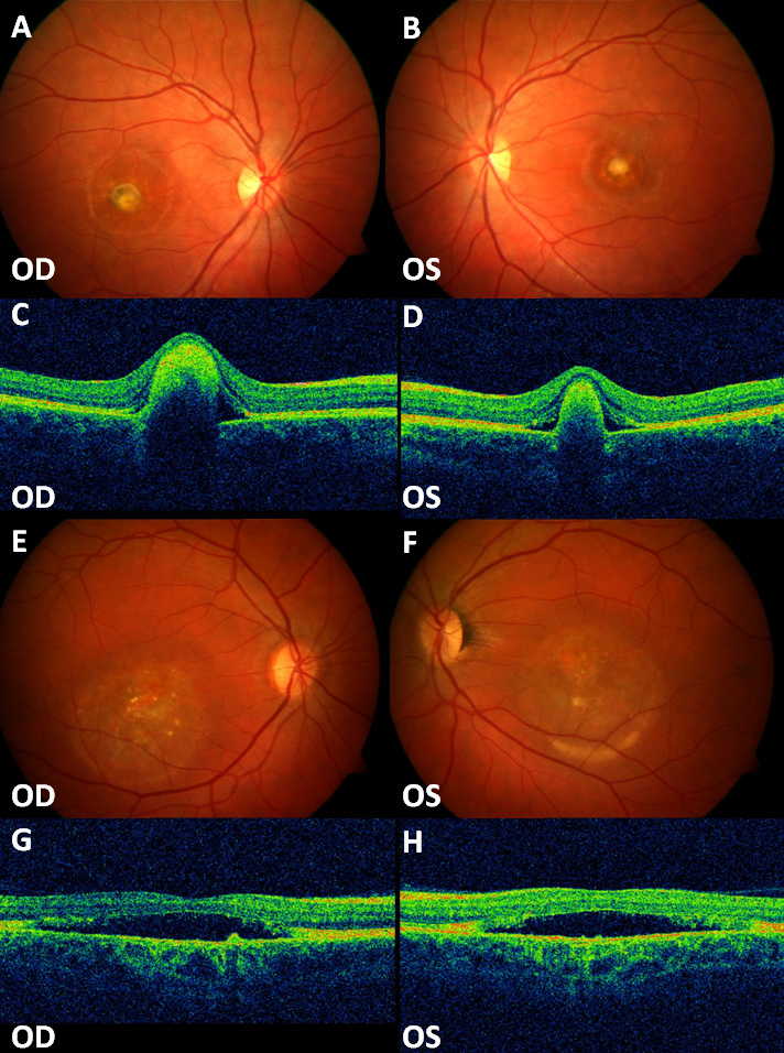
 Figure 4 of
Sodi, Mol Vis 2012; 18:2736-2748.
Figure 4 of
Sodi, Mol Vis 2012; 18:2736-2748.