Figure 3. Fundus photographs, OCT and EOG of Patient M-II-1 carrying the novel sequence variant p.Ile295del. A, B: Vitelliform lesion with partial re-absorption of the vitelliform material. C, D: Macular detachment of the neurosensory retina partially occupied by hyperreflective material. E, F: Reduced Arden Test. OD represents the right eye, OS represents the left eye.
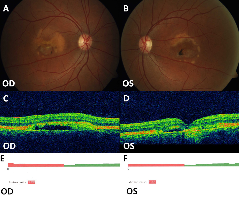
 Figure 3 of
Sodi, Mol Vis 2012; 18:2736-2748.
Figure 3 of
Sodi, Mol Vis 2012; 18:2736-2748.