Figure 1. Different stages of VMD (Fundus photographs and OCT). A, B: Vitellifom disc (Patient R-II-1). C, D: Vitelliruptive stage (Patient C-II-1). E, F: Pseudohypopyon stage (Patient B-II-1). G, H: Macular atrophy (Patient Q-I:-1). I, J: Macular fibrosis (Patient I-II-1). OD represents the right eye, OS represents the left eye.
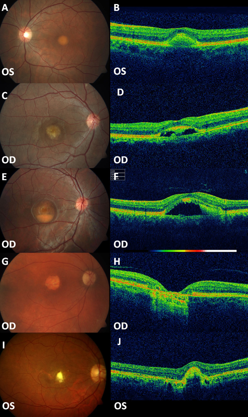
 Figure 1 of
Sodi, Mol Vis 2012; 18:2736-2748.
Figure 1 of
Sodi, Mol Vis 2012; 18:2736-2748.