Figure 3. Detection of herpes simplex virus 1 DNA in the trigeminal ganglia. Five left trigeminal ganglia (TG) from each treatment group
were pooled and processed for the detection of herpes simplex virus 1 (HSV-1) DNA by PCR. The tissues were homogenized, DNA
extracted, and amplified using real-time PCR. HSV-1 DNA (at a threshold cycle, CT, of around 15) was detected in trigeminal
tissues derived from KOS-tk12 and KOS(Rid1)-tk12 inoculated animals. CT values represent the fractional cycle number at which
fluorescence passes a fixed threshold (indicated). The tissues from mock-infected animals were negative.
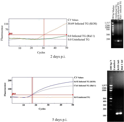
 Figure 3 of
Shukla, Mol Vis 2012; 18:2711-2716.
Figure 3 of
Shukla, Mol Vis 2012; 18:2711-2716.