Figure 4 of
Kumar, Mol Vis 2012; 18:2687-2699.
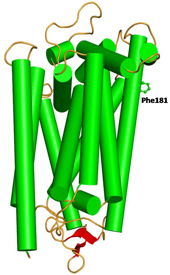
Figure 4.
Model structure of wild-type human mitochondrial cytochrome b in cartoon rendering indicating secondary structure: helices (green), β-strand (red), and loop (orange). The side chain of Phe181 is shown as a ball and stick.
 Figure 4 of
Kumar, Mol Vis 2012; 18:2687-2699.
Figure 4 of
Kumar, Mol Vis 2012; 18:2687-2699.