Figure 2. Chemical and genetic fractionation of the retina. A: Juxtaposed images of histological sections comparing the retinas of a normal (control) mouse, with one whose inner retina
has been damaged by glutamate, and another that was taken from a C3H mouse suffering the loss of the distal retina. B: The concentrations of five amino acids in each preparation. The latter values represent the averages from six different groups
of dark-adapted animals. (Modified from Cohen et al., 1973, with the permission of the publishers).
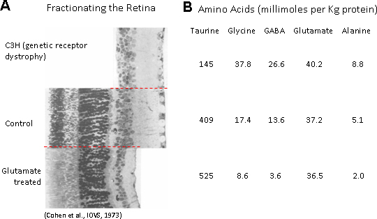
 Figure 2 of
Ripps, Mol Vis 2012; 18:2673-2686.
Figure 2 of
Ripps, Mol Vis 2012; 18:2673-2686.