Figure 8. Retinal ganglion cell axon restoration and optic nerve regeneration in retinal progenitor cell (RPC)-transplanted eyes. A, B: Cranial images from tamoxifen-treated control- (A) and RPC-injected (B) mice sixteen weeks after transplantation. C-E: Flat-mounted retinas from control (C) noninjected (D) and injected (E) eyes with anti-Pou4f2 (green) and anti-NFL (red) antibodies. Dashed boxes depict regions at higher magnification in panels
F-H: Arrows, optic nerve; asterisk, RPC-transplanted eye, OHN, optic nerve head. Scale bars: A, B: 3 mm; C-E: 200 μm, F-H: 50 μm.
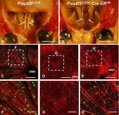
 Figure 8 of
Cho, Mol Vis 2012; 18:2658-2672.
Figure 8 of
Cho, Mol Vis 2012; 18:2658-2672.