Figure 6. Transplanted retinal progenitor cells differentiate into retinal ganglion cells (RGCs). A-F: Retinal sections from six (A-C) and ten weeks (G-I) after transplantation were immunostained with anti-Pou4f2 (green) and anti-NFL (red) antibodies. Sections were counterstained
with DAPI. Dashed boxes indicate regions depicted at higher magnification in panels D-F and J-L. Flat-mounted retinas at sixteen weeks after transplantation were immunostained with anti-Pou4f2 (green) and NFL (red) antibodies
(M-O). Arrowheads, Pou4f2-expressing RGCs. ONL, outer nuclear layer; INL, inner nuclear layer; GCL, ganglion cell layer; ONH,
optic nerve head. Scale bars: 100 μm.
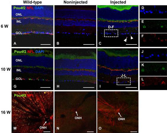
 Figure 6 of
Cho, Mol Vis 2012; 18:2658-2672.
Figure 6 of
Cho, Mol Vis 2012; 18:2658-2672.