Figure 5. Bromodeoxyuridin (BrdU) pulse-chase-labeled retinal progenitor cells (RPCs) in transplanted RPCs. Retinal sections of wild-type
(A), noninjected (B), and injected eyes (C, D) were immunostained with anti-BrdU antibody at three or six weeks after transplantation (brown). Dashed boxes indicate regions
depicted at higher magnification in panels E-H: Arrowheads represent BrdU-positive cells that have incorporated into the ganglion cell layer (GCL), and arrows represent
BrdU-positive cells that have not incorporated into the GCL (brown). Sections were counterstained with hematoxylin. Scale
bars: A-D: 100 μm; E-H: 50 μm.
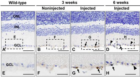
 Figure 5 of
Cho, Mol Vis 2012; 18:2658-2672.
Figure 5 of
Cho, Mol Vis 2012; 18:2658-2672.