Figure 2. Green fluorescent protein (GFP)-expressing retinal progenitor cells at two to three weeks after intravitreal transplantation.
A, B: Retinal sections from noninjected and injected retinas under a GFP filter with propidium iodide (PI) counterstaining (red).
Dotted boxes indicate regions depicted at higher magnification in panels C-F. G-J: Retinal sections were immunostained with anti-GFP (green) and anti-Pou4f2 (red) antibodies. Cells in panel G were counterstained with DAPI (blue). Dashed boxes indicate regions depicted at higher magnification on each upper-right
corner. ONL, outer nuclear layer; INL, inner nuclear layer; GCL, ganglion cell layer. Scale bars: A, B: 100 μm; C-F: 50 μm; G-J: 100 μm.
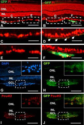
 Figure 2 of
Cho, Mol Vis 2012; 18:2658-2672.
Figure 2 of
Cho, Mol Vis 2012; 18:2658-2672.