Figure 10. Green fluorescent protein (GFP)- and Pou4f2-expression in transplanted cells six weeks after intravitreal injection. Retinal
flat mounts with DAPI (A), anti-GFP antibody (B) anti-Pou4f2 antibody (C): and merged imaged with panel B and C (D). Arrows indicate transplanted cells. ONH, optic nerve head. Scale bars: 50 μm.
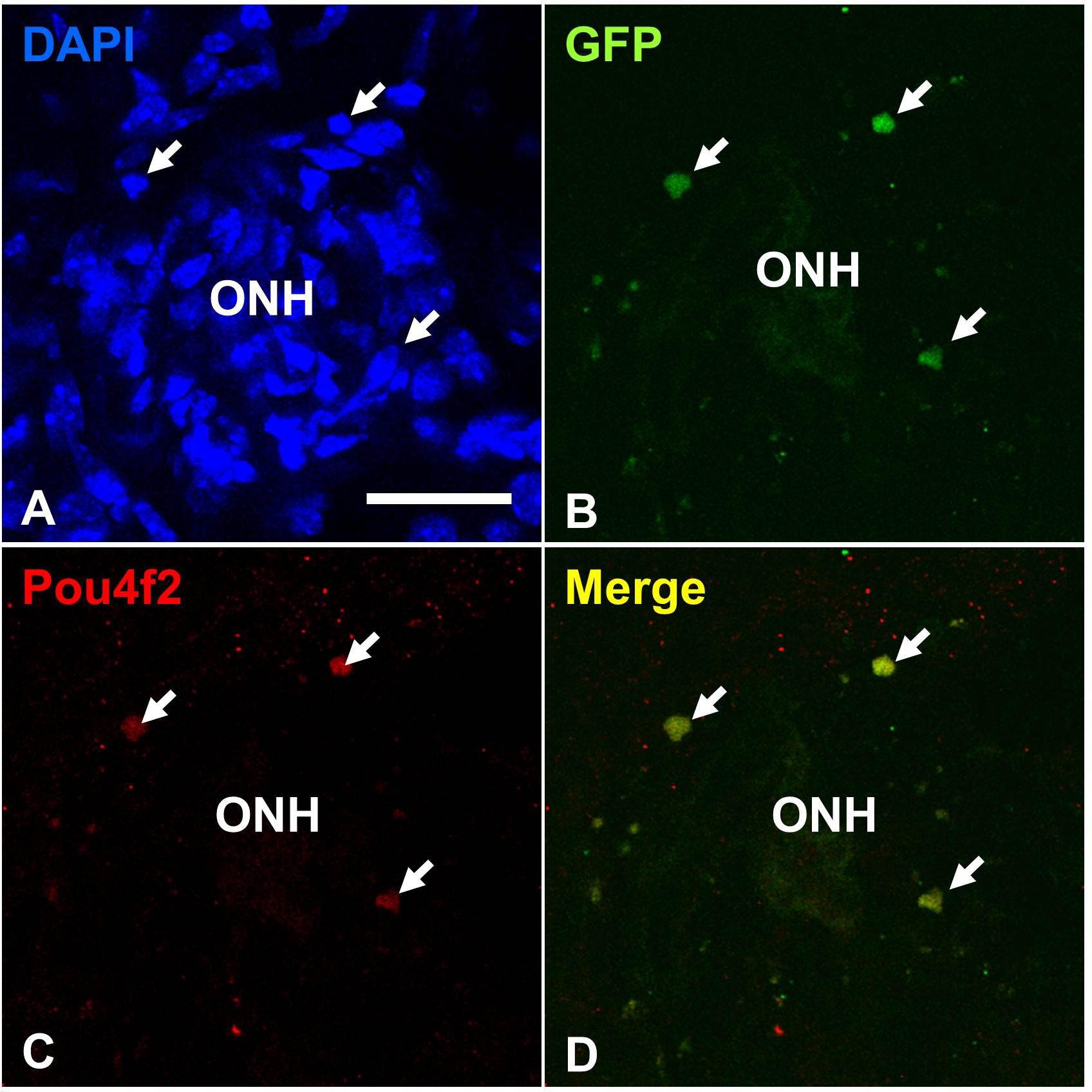
 Figure 10 of
Cho, Mol Vis 2012; 18:2658-2672.
Figure 10 of
Cho, Mol Vis 2012; 18:2658-2672.