Figure 5. Immunolocalizations of amyloid β1–4 (Aβ1–42) in the optic nerve head of cynomolgus monkeys. Representative photographs showing of amyloid β1–4 (Aβ1–42)/glial fibrillary acidic protein (GFAP: A). Aβ1–42/phosphorylated neurofilament H (SMI-31: B) and Aβ1–42/non-phosphorylated neurofilament H (SMI-32: C) immunofluorescence stainings from monkey optic nerve head (ONH) at 11 weeks after laser photocoagulation treatment. Aβ1–42 was co-localized with GFAP but not SMI-31 or SMI-32 in the ONH at 11 weeks after laser photocoagulation treatment. Horizontal
scale bars indicate 30 µm.
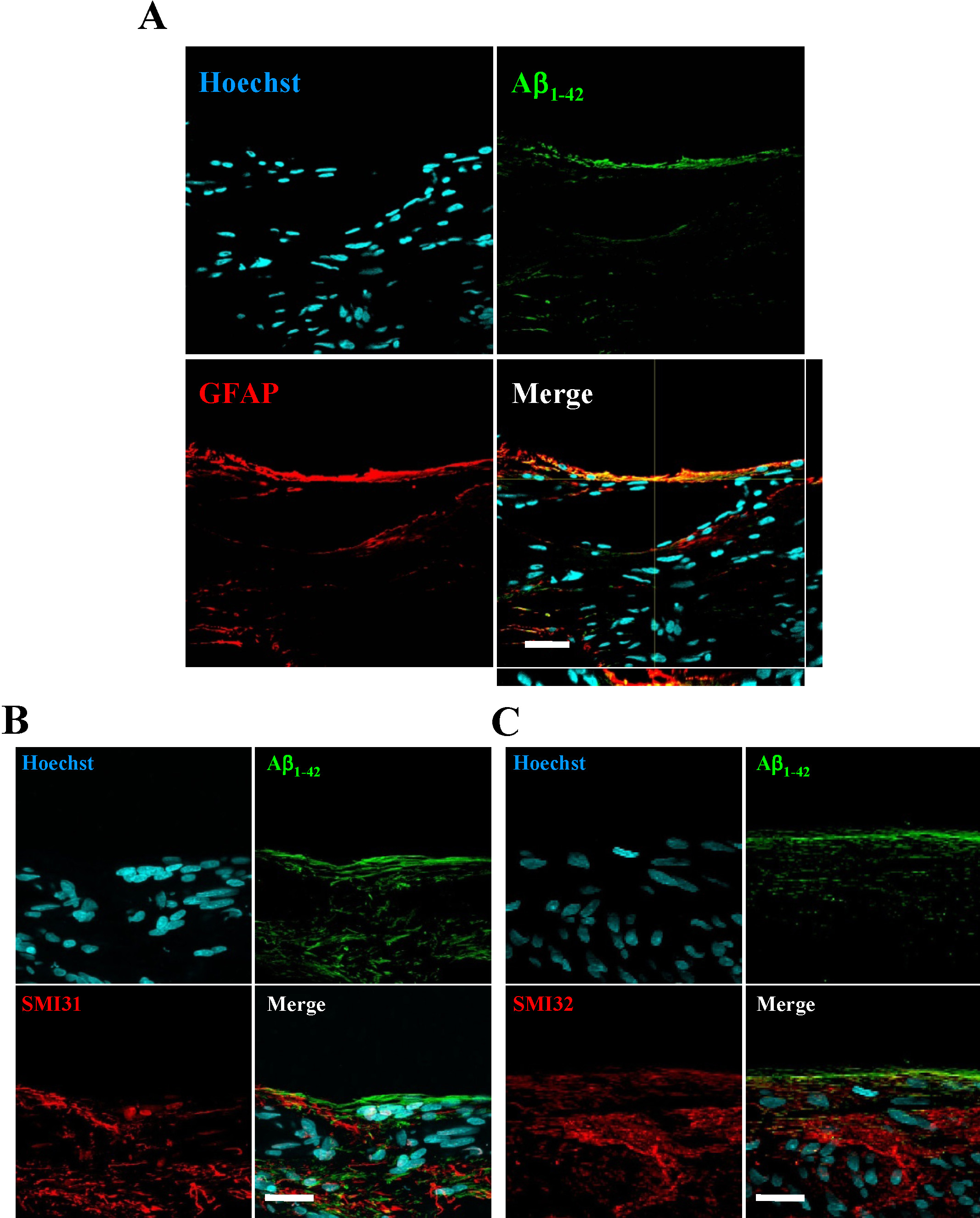
 Figure 5 of
Ito, Mol Vis 2012; 18:2647-2657.
Figure 5 of
Ito, Mol Vis 2012; 18:2647-2657.