Figure 4. Representative photographs show amyloid β1–42 (Aβ1–42)/glial fibrillary acidic protein (GFAP; A), Aβ1–42/phosphorylated neurofilament H (SMI-31; B) and Aβ1–42/non-phosphorylated neurofilament H (SMI-32; C) immunofluorescence stainings from monkey retina at 11 weeks after the laser photocoagulation treatment. Aβ1–42 was colocalized with GFAP, but not SMI-31- or SMI-32, in the nerve fiber layer (NFL) and the ganglion cell layer (GCL) of
the retina at 11 weeks after the laser photocoagulation treatment. Abbreviations are as follows: Hoechst represents Hoechst33342;
IPL represents inner plexiform layer; INL represents inner nuclear layer. Horizontal scale bars indicate 30 µm.
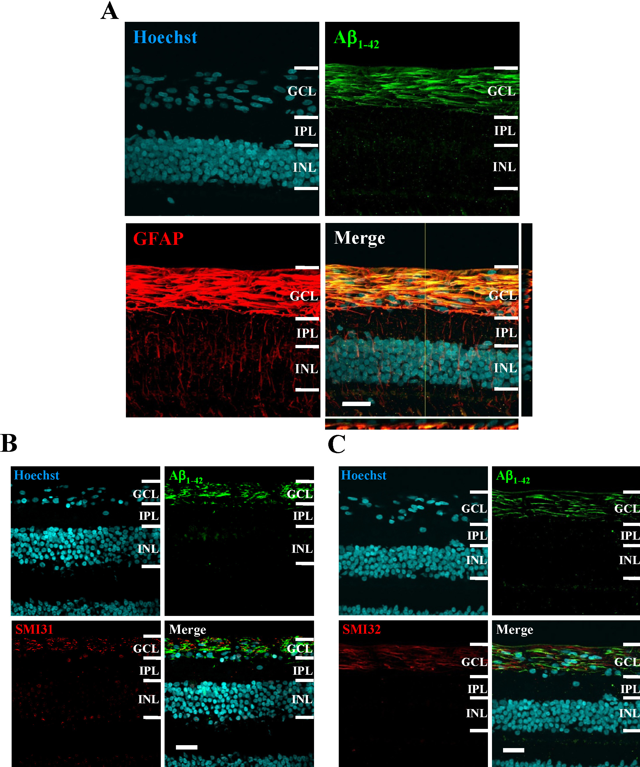
 Figure 4 of
Ito, Mol Vis 2012; 18:2647-2657.
Figure 4 of
Ito, Mol Vis 2012; 18:2647-2657.