Figure 2. Time-dependent changes in the expression of amyloid β1–42 in retina and optic nerve head after laser photocoagulation treatment in cynomolgus monkeys. Representative photographs of
retinal and optic nerve head (ONH) sections immunostained with anti-amyloid β (Aβ1–42, green) and counterstained with Hoechst 33,342 (blue) were obtained from the monkeys that had the pressure in their left
eye elevated at 4 to 24 weeks. Aβ1–42 immunoreactivity was found to be increased in the nerve fiber layer (NFL), the ganglion cell layer (GCL), and ONH of the
glaucomatous left eye from 11 weeks after IOP elevation, while there was no Aβ1–42 immunoreactivity in the non-treated eye. Abbreviations are as follows: IOP represents intraocular pressure; IPL represents
inner plexiform layer; INL represents inner nuclear layer; OPL represents outer plexiform layer. Horizontal scale bars indicate
50 µm.
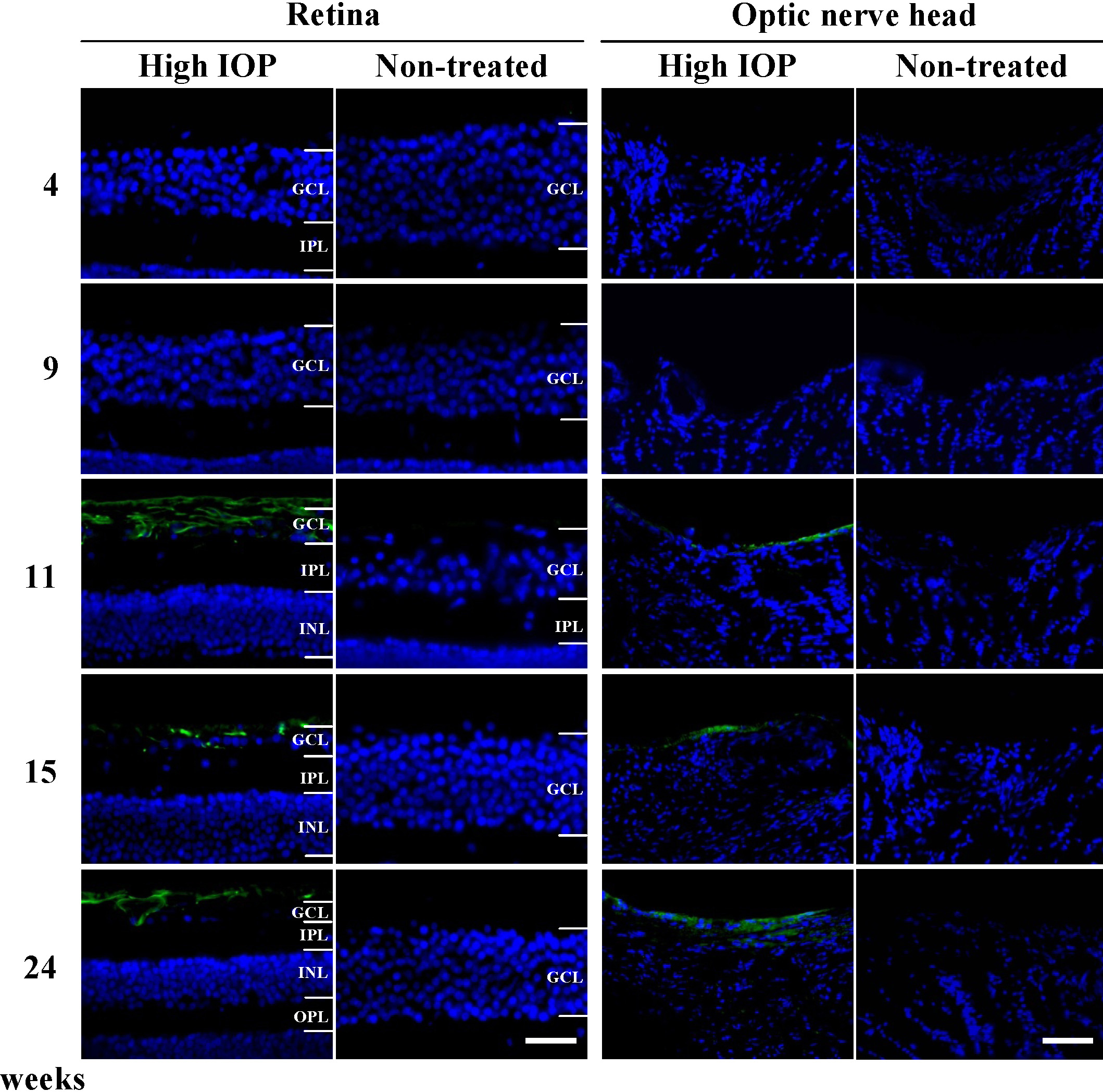
 Figure 2 of
Ito, Mol Vis 2012; 18:2647-2657.
Figure 2 of
Ito, Mol Vis 2012; 18:2647-2657.