Figure 1. Morphological changes in retina and optic nerve head after chronic elevation of intraocular pressure. Representative photographs
A: shows hematoxylin and eosin staining sections of retina and optic nerve head obtained from the eyes that had the pressure
in their left eye elevated for 4 and 15 weeks and nontreated eye. Each scale bar indicates 100 μm for retina and 250 μm for
ONH. Abbreviations are as follows: IOP represents intraocular pressure; GCL represents ganglion cell layer; IPL represents
inner plexiform layer; INL represents inner nuclear layer; OPL represents outer plexiform layer. Fundus photographs in monkey.
Representative fundus photographs showing nontreated eye, and treated eye at 133 weeks after IOP elevation B.
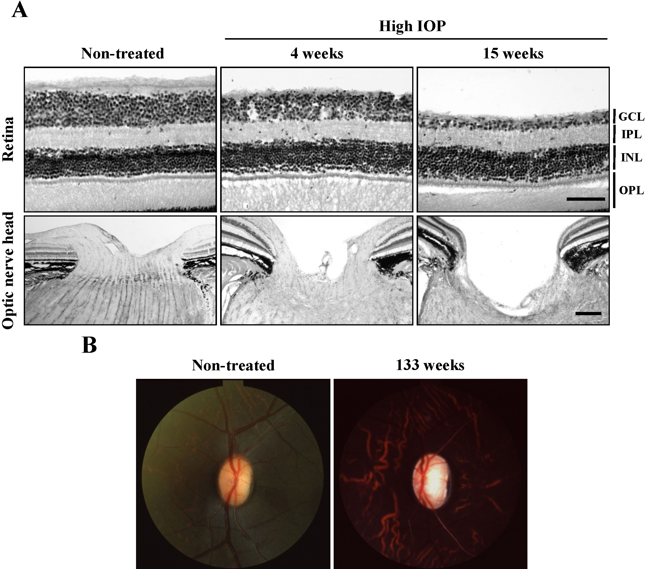
 Figure 1 of
Ito, Mol Vis 2012; 18:2647-2657.
Figure 1 of
Ito, Mol Vis 2012; 18:2647-2657.