Figure 1. Analysis of the anterior segment of the patient's eyes at presentation with spectral-domain anterior segment optical coherence
tomography. The images are automatically corrected with the anterior segment optical coherence tomography (AS-OCT) software
accounting for the optical effect of the anterior and posterior surfaces of the cornea. On the left: horizontal B-scans of
the anterior chamber. CCT: central corneal thickness; ACD: anterior chamber depth, ACD [epi]: distance from the corneal epithelium
to the lens anterior surface; ACD [endo]: distance from the corneal endothelium to the lens anterior surface; AR1-AR2: anterior
chamber width, from temporal to nasal angular recess. On the right: vertical B-scans centered on the lens. Lens thickness
is measured along the fixation axis.
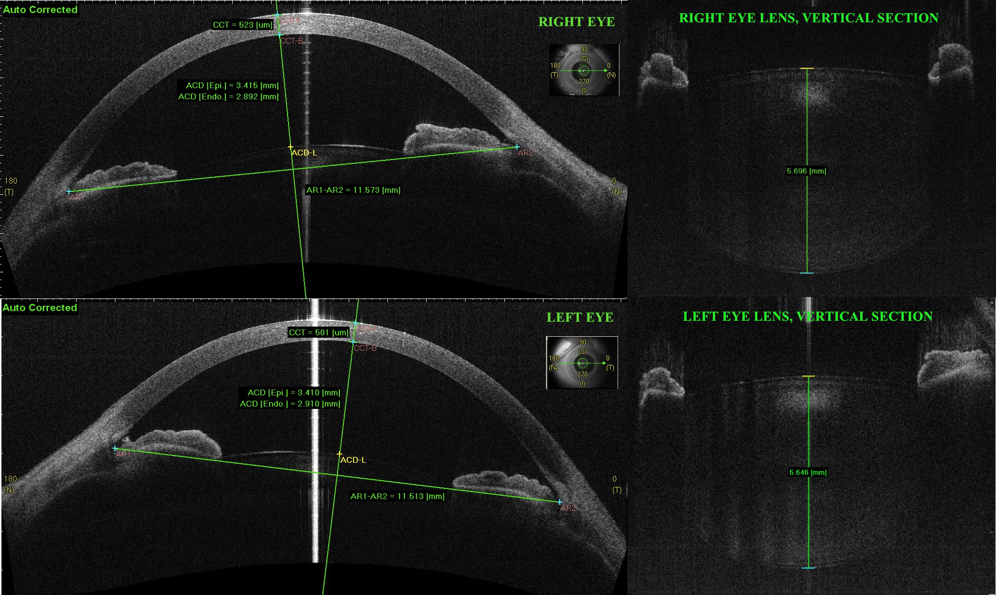
 Figure 1 of
Neri, Mol Vis 2012; 18:2623-2632.
Figure 1 of
Neri, Mol Vis 2012; 18:2623-2632.