Figure 3. Quantification of αSMA mRNA. A: and protein B: levels in ±TGFβ1 treated HCF cultures showing anti-fibrotic response of sTGFβRII-Fc gene transfer. A: Real-time PCR showed that TGFβ1 treatment caused 5±1.4 fold increase in αSMA mRNA (#; p<0.01 than TGFβ1 untreated) and nanoparticle-mediated
sTGFβRII-Fc transfection significantly lowered αSMA mRNA (0.36±0.08 fold, ψ; p<0.01 compared to TGFβ1-treated and ϕ; p<0.05
compared to lipofectamine-transfected). B: In western blotting an expected significant increase in αSMA levels were detected in TGFβ1-treated HCF over the TGFβ1-untreated
HCF (#; p<0.001). Nanoparticles-mediated sTGFβRII-Fc gene transfer into HCF demonstrated a significant 96±3% decrease in αSMA
levels (ψ; p<0.001 compared to TGFβ1-treated and ϕ; p<0.01 compared to lipofectamine-transfected). Lipofectamine-delivered
sTGFβRII-Fc HCF used for comparison showed less than 60% decrease in αSMA expression.
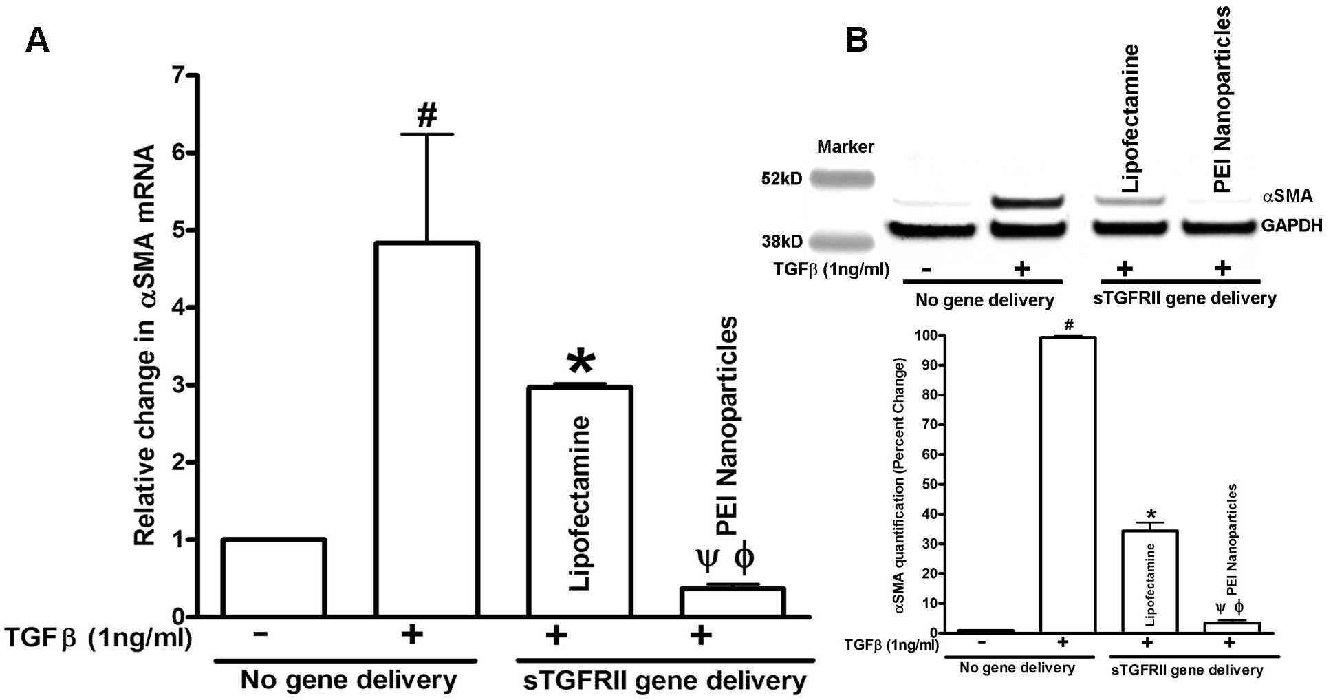
 Figure 3 of
Sharma, Mol Vis 2012; 18:2598-2607.
Figure 3 of
Sharma, Mol Vis 2012; 18:2598-2607.