Figure 5. Effect of IMD-0354 on phosphorylated inhibitors of κB (green) in the iris-ciliary body 3 h after lipopolysaccharide injection.
No phosphorylated inhibitor of κB (p-IκB signal was detected in negative control A: where no p-IκB antibodies were applied. The p-IκB signals were similarly expressed in cytoplasm of naïve controls B: and IMD-0354-treated (30 mg/kg) LPS injected rats C: Intensive p-IκB expression was observed in cytoplasm of iris-ciliary body (ICB) cells in untreated endotoxin-induced uveitis
(EIU) rats D: Cell nuclei were stained with 4',6-diamidino-2-phenylindole (DAPI) as blue.
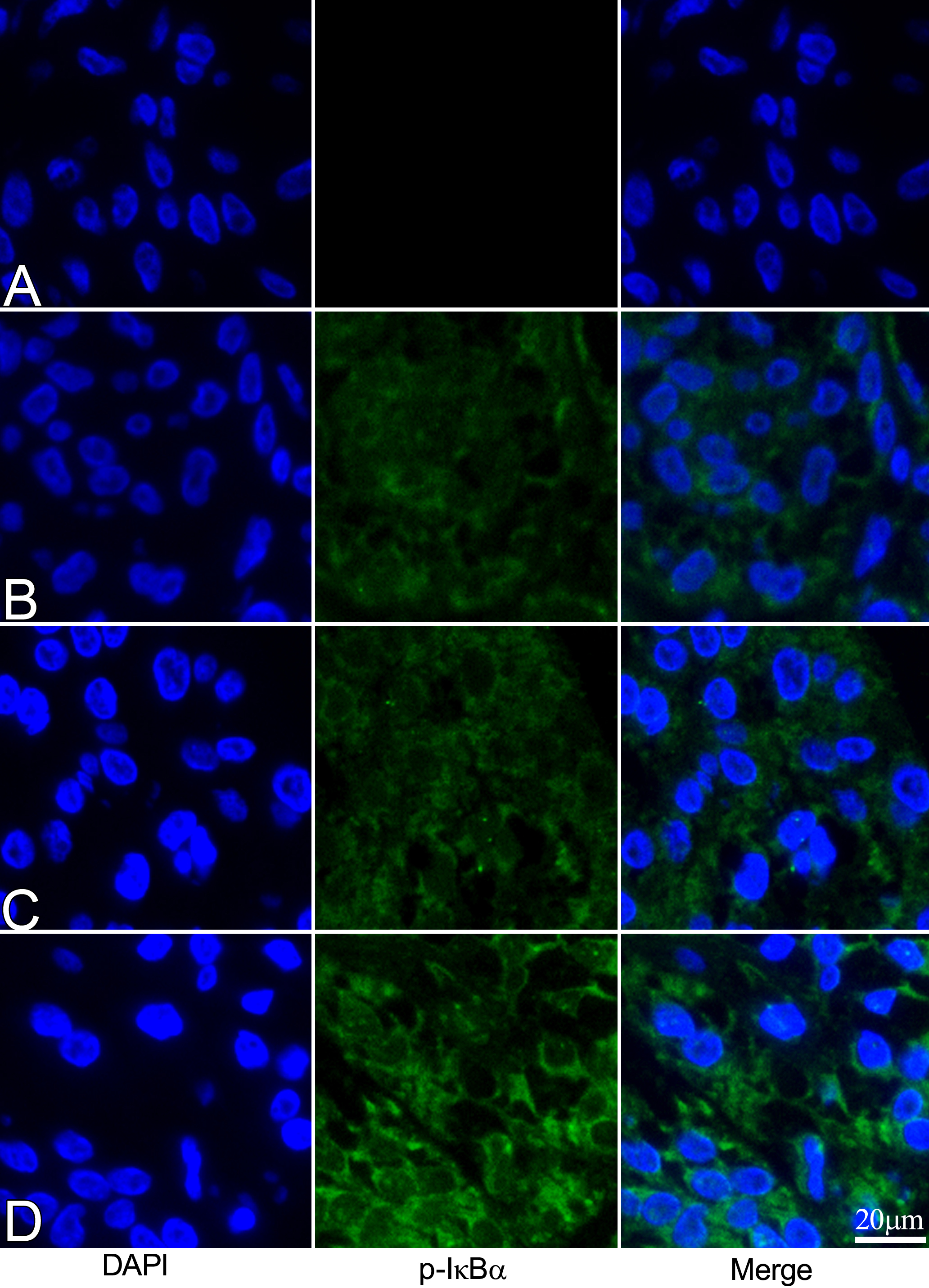
 Figure 5 of
Lennikov, Mol Vis 2012; 18:2586-2597.
Figure 5 of
Lennikov, Mol Vis 2012; 18:2586-2597.