Figure 1. Histological changes in the
iris-ciliary body, vitreous cavity, and retina 24 h after
lipopolysaccharide injection. Photographs on the left side show
the iris-ciliary body (ICB) region, and those on the right side
show the vitreous and retina in rats. Control animals E,
J: were not injected with lipopolysaccharide (LPS); no
inflammation was observed. Severe inflammatory cell infiltration
was observed in endotoxin-induced uveitis (EIU) rats D:
I: In the group of EIU rats treated with IMD-0354 (30
mg/kg) A, F: 10 mg/kg; B, G:
reductions in cell infiltration were observed compared to
untreated EIU rats. No noticeable reduction in cell infiltration
was observed in EIU rats treated with IMD-0354 (3 mg/kg) C:
H: Infiltrating cells in the ICB of sections K:
and in the posterior part of the eye (l) were counted and
averaged. Data are shown as mean±standard error of mean (SEM;
n=8). *p<0.05, **p<0.01, significantly different from the
LPS group.
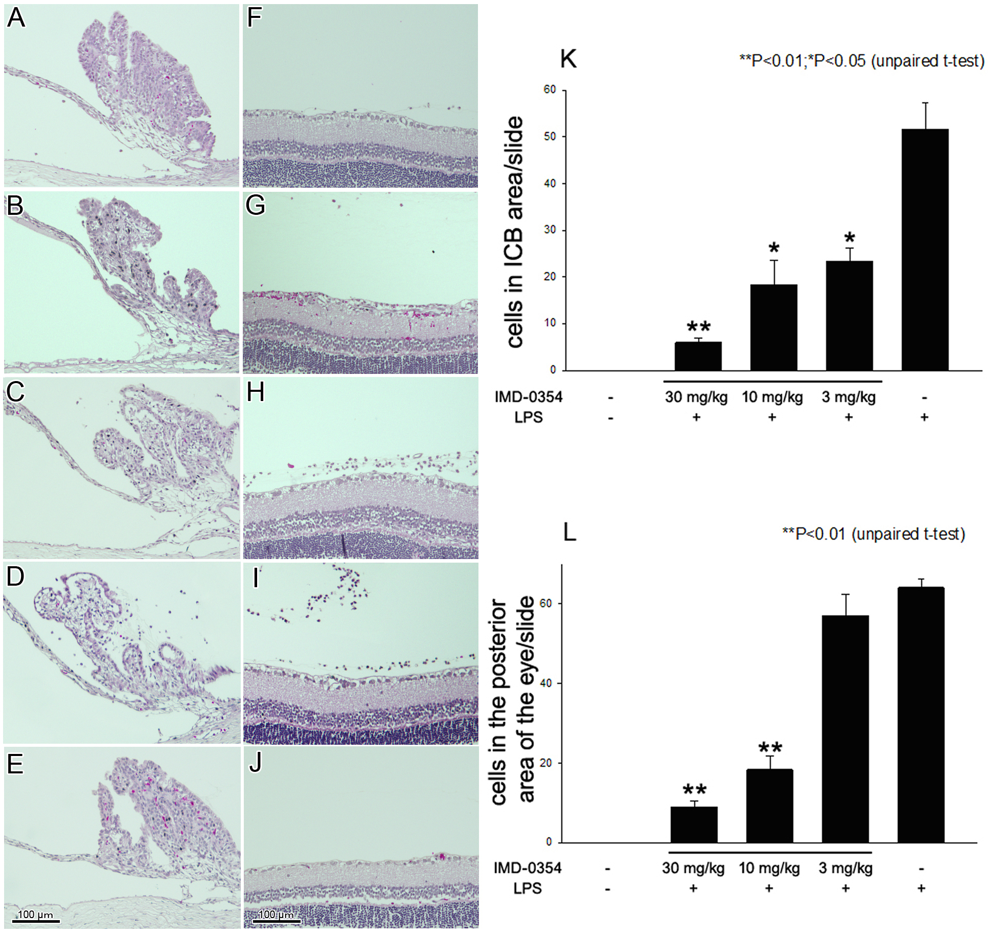
 Figure 1
of Lennikov, Mol Vis 2012; 18:2586-2597.
Figure 1
of Lennikov, Mol Vis 2012; 18:2586-2597.