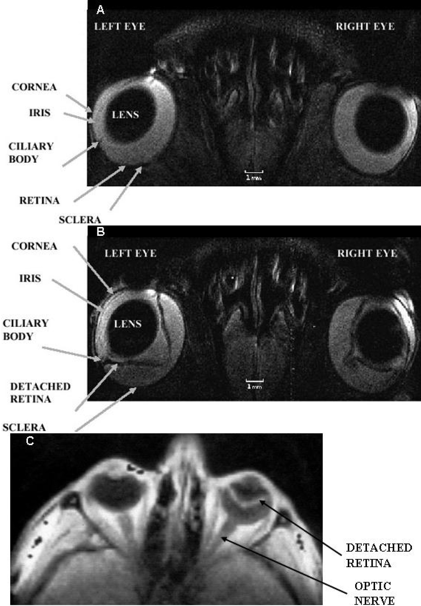
Figure 2. Selected regions of in vivo
MRI images of the rat (
A and
B and the human
C
eye. Eye structures such as the lens, cornea, iris, ciliary
body, retina and sclera are visualized. In
A both rat
eyes are normal. The left rat eye in
B is at peak of
experimental autoimmune uveitis and the right rat eye in the
same figure shows post-peak of disease. The spatial resolution
of each MRI rat image was 60×60×700 μm
3 and the
acquisition time was 30 min. The left human eye in
C:
shows no retinal detachment, while the right human eye in the
same figure shows a large retinal detachment. The MRI images in
A and
B are T
2-weighted, while that in
C is T
1-weighted. The images in
A and
B were reproduced from [
38]
while that in
C: from [
43]
with permissions from John Wiley & Sons and Elsevier Ltd
respectively.
 Figure 2
of Fanea, Mol Vis 2012; 18:2538-2560.
Figure 2
of Fanea, Mol Vis 2012; 18:2538-2560.