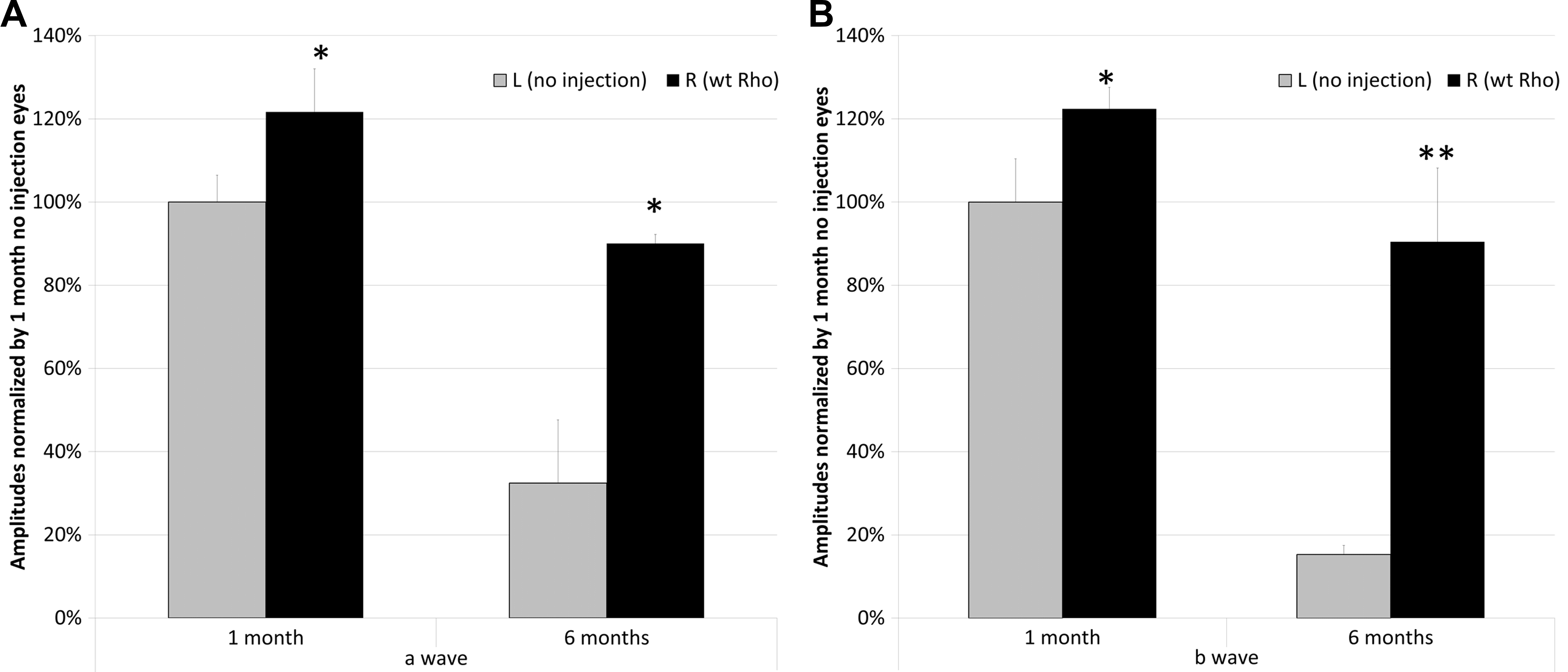
Figure 2. Improvement of
electroretinography (ERG) response by single AAV injection of
normal mouse rhodopsin cDNA (WT
Rho) in P23H transgenic
mice [
89].
Bars represent the average of five scotopic ERG scans at 0 dB
(2.6 cd (cd)-s/m
2) a-wave response.
A:
and b-wave response.
B: at 1 month and 6 months post
injection. ERG amplitudes of 1 month uninjected P23H eyes were
set as 100%.
A: Compared with that of corresponding
contralateral eyes, injection of AAV-
Rho demonstrated a
significant increase in a-wave amplitudes at both 1 month (122%)
and 6 months (90%) time points. (*p<0.05, n=6).
B:
Compared with contralateral eyes, injection of WT
Rho
demonstrated the same significant increase in b-wave amplitudes
as that of a-wave response at both 1 month (122%) and 6 months
(90%) time points. (*p<0.05, ** p<0.005, n=6). Although
injection injury can induce protective cytokines such as CNTF,
this effect peaks within a few days of injection and is complete
before the first measurements were made.
 Figure 2
of Rossmiller, Mol Vis 2012; 18:2479-2496.
Figure 2
of Rossmiller, Mol Vis 2012; 18:2479-2496.