Figure 3. Images of patient 2. Fundus
autofluorescence (FAF) images C and D correspond
with the white square regions in images A and B,
respectively. The horizontal white dashed lines represent the
position of the spectral domain-optical coherence tomography
(SD-OCT) images E and F on the corresponding en
face image. FAF revealed widespread retinal disease. Relative
foveal sparing was present bilaterally as evidenced by a uniform
autofluorescence pattern at the foveae (black arrows). The
corresponding SD-OCT images revealed relative preservation of
the inner segment–outer segment junctions of the photoreceptors
(IS/OS), external limiting membrane (ELM), and outer nuclear
layer (ONL) in this region. The horizontal borders of the
regions with preserved IS/OS are indicated by the vertical white
dashed lines on the corresponding FAF and SD-OCT images. Outside
these regions, absence of IS/OS was associated with functionally
preserved retinal pigment epithelium (RPE), at least as
identified by FAF (C, D). Qualitatively normal
thickness RPE was also observed in regions with loss of IS/OS (F,
white arrow).Corresponding size bars for A and B,
C and D, and E and F are included
in A, C and E, respectively
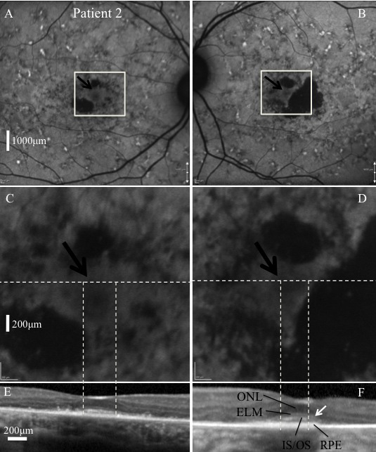
 Figure 3
of Burke, Mol Vis 2012; 18:227-233.
Figure 3
of Burke, Mol Vis 2012; 18:227-233.