Figure 7. RGC images expressing cyan fluoresceint protein (CFP) in the retina. Whole mount retinal images of control eye A: and laser-treated eye B: were taken under the fluorescein microscope. Magnified images corresponding to the white box in A and B: at 1200 μm from the optic disc in the superior area are C: and D: The numbers of RGC expressing CFP decreased in the laser-treated eye. Scale bar is 50 μm.
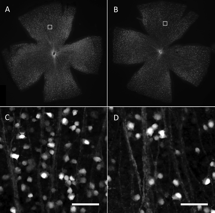
 Figure 7 of
Tsuruga, Mol Vis 2012; 18:2468-2478.
Figure 7 of
Tsuruga, Mol Vis 2012; 18:2468-2478.