Figure 3. Proliferation of B16F10
cells after bevacizumab treatment. Bevacizumab was added to
B16F10 cells in vitro under normoxic (top) and hypoxic (bottom)
circumstances and proliferation was measured by WST-1 assay on
days one, two and three after addition of bevacizumab. Cell
density is expressed in absorbance (optical density, OD). A high
dose of bevacizumab inhibited cell growth under hypoxic
conditions.
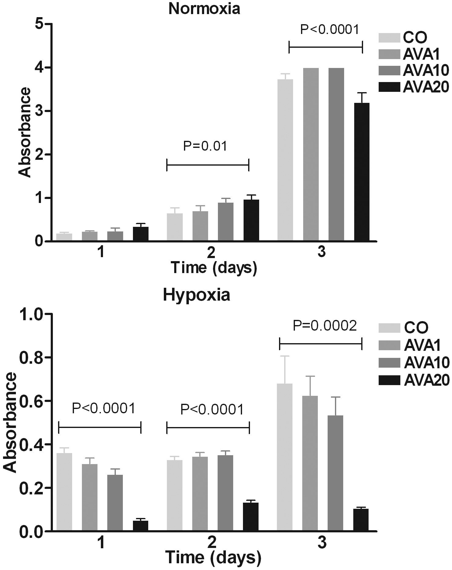
 Figure 3
of el Filali, Mol Vis 2012; 18:2454-2467.
Figure 3
of el Filali, Mol Vis 2012; 18:2454-2467.