Figure 1. Ophthalmological images of two siblings affected by retinitis pigmentosa, nanophthalmus and optic disc drusen. Fundus photography
of the right eye of patient IV:2 A: and of patient IV:3 B: showed atrophy of the retina outside the fovea and spots of hyperpigmentation. B-mode ultrasound of the left eye of patient
IV:2 C: revealed optic disc drusen (indicated with the yellow arrow). Optical coherence tomography scan of the macula of patient
IV:2 showed intraretinal edema and atrophy of the outer retinal layers D.
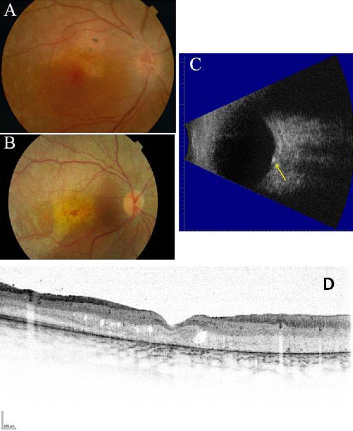
 Figure 1 of
Paun, Mol Vis 2012; 18:2447-2453.
Figure 1 of
Paun, Mol Vis 2012; 18:2447-2453.